Yonsei Med J.
2005 Jun;46(3):368-371. 10.3349/ymj.2005.46.3.368.
Contrast Sensitivity Function of Sound Eye after Occlusion Therapy in the Amblyopic Children
- Affiliations
-
- 1Department of Ophthalmology, CHA University Medical College, Kyunggi, Korea.
- 2Department of Ophthalmology, Yonsei University and Institute of Vision Research, Seoul, Korea. shhan222@yumc.yonsei.ac.kr
- KMID: 1734071
- DOI: http://doi.org/10.3349/ymj.2005.46.3.368
Abstract
- To verify the changes of mesopic and photopic contrast sensitivity function of sound eye whose visual acuity was kept the same after occlusion therapy in the amblyopic children. Fourteen sound eyes of amblyopic children (mean; 7.67 years; S.D., 1.50 years) who kept their visual acuity the same after the occlusion therapy were tested. The children had 6 hours of part-time patch therapy for 3 months prior to this examination. Among 14 amblyopic children, 8 were anisometric and 6 were strabismic amblyopes. Using the visual capacity analyzer which measures the minimal contrast level at from low to high spatial frequencies, the contrast sensitivity of sound eye was measured, under both photopic and mesopic condition, before and after 3 months of occlusion therapy. Comparing the contrast sensitivity of sound eye after the occlusion therapy to that before the occlusion, there was no statistical difference in photopic condition. When it comes to mesopic condition, the contrast sensitivity decreased at the intermediate spatial frequency level (3-13 c.p.d, p=0.028) after the occlusion therapy. The occlusion caused statistically significant decrease in mesopic contrast sensitivity, when the visual acuity was not changed after the occlusion therapy. It may indicate that mesopic contrast sensitivity can be considered as a useful tool for early detection of hidden occlusion amblyopia.
MeSH Terms
Figure
-
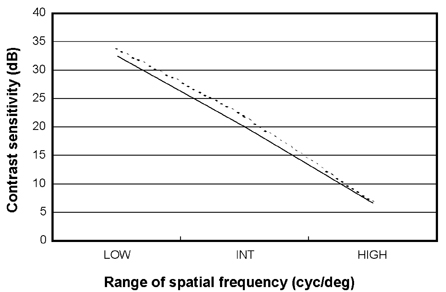
Fig. 1 Average contrast sensitivity plotted as a function of spatial frequency under photopic condition (The broken line: before occlusion, the solid line: after 3 months of occlusion).
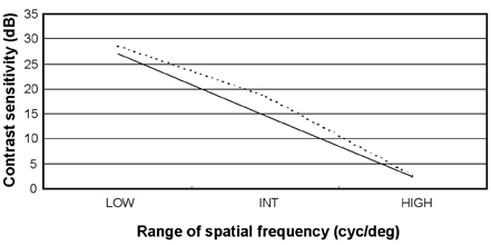
Fig. 2 Average contrast sensitivity plotted as a function of spatial frequency under mesopic condition (The broken line: before occlusion, the solid line: after 3 months of occlusion).
Cited by 1 articles
-
Comparison of Higher-Order Aberration and Contrast Sensitivity in Monofocal and Multifocal Intraocular Lenses
Chang Yeom Kim, So-Hyang Chung, Tae-im Kim, Young Jae Cho, Geunyoung Yoon, Kyoung Yul Seo
Yonsei Med J. 2007;48(4):627-633. doi: 10.3349/ymj.2007.48.4.627.
Reference
-
1. Zimmern RL, Campbell FW, Wilkinson IM. Subtle disturbances of vision after optic neuritis elicited by studying contrast sensitivity. J Neurol Neurosurg Psychiatry. 1979. 42:407–412.2. Hyvarinen L. Contrast sensitivity in visually impaired children. Acta Opthalmol. 1983. 157:Suppl. 58–62.3. Volkers ACW, Hagemans KH, Vanderwildt GJ, Schimitz PIM. Spatial contrast and the diagnosis of amblyopia. Br J Ophthalmol. 1987. 71:58–65.4. Rydberg A, Han Y. Assessment of contrast sensitivity in children aged 3 years 9 months-6 years with normal vision, visual impairment due to ocular disease and strabismic amblyopia. Strabismus. 1999. 7:79–95.5. Woo GC, Dalziel CC. A pilot study of contrast sensitivity assessment of the CAM treatment of amblyopia. Acta Ophthalmol (Copenh). 1981. 59:35–37.6. Lew H, Seong GJ, Kim SK, Lee JB, Han SH. Mesopic contrast sensitivity function in amblyopic children. Yonsei Med J. 2003. 44:995–1000.7. Howell ER, Mitchell DE, Keith CG. Contrast thresholds for sine gratings of children with amblyopia. Invest Ophthalmol Vis Sci. 1983. 24:782–787.8. Rogers GL, Bremer DL, Leguire LE. The contrast sensitivity Function and childhood amblyopia. Am J Ophthalmol. 1987. 104:64–68.9. Higgins KE, Daugman JG, Mansfield RJ. Amblyopic contrast sensitivity: insensitivity to unsteady fixation. Invest Ophthalmol Vis Sci. 1982. 23:113–120.10. Kubota N, Usui C. The development of occlusion amblyopia following atropine therapy for strabismic amblyopia. Nippon Ganka Gakkai Zasshi. 1993. 97:763–768.11. Pardhan S, Gilchrist J. Binocular contrast summation and inhibition in amblyopia. Doc Ophthalmologica. 1992. 82:239–248.
- Full Text Links
-
- Actions
-
Cited
- CITED
-
- Close
- Share
-
- Similar articles
-
- The Change of Contrast Sensitivity in Amblyopic Patient after Occlusion Therapy using ACV
- Mesopic Contrast Sensitivity Functions in Amblyopic Children
- The Efficacy of Intermittent Atropine Penalization in Amblyopic Children Who Have Failed Patching Therapy
- The Change of Pupil Cycle Time after Occlusion Therapy in Amblyopia
- Clinical Analysis of Successfully Treated Amblyopia with Anisometropia, Strabismis, and Combined Cause
