Intravascular Papillary Endothelial Hyperplasia (Masson's Hemangioma) of the Liver: A New Hepatic Lesion
- Affiliations
-
- 1Department of Surgery, St. Vincent's Hospital, The Catholic University of Korea, Suwon, Korea. hchin@catholic.ac.kr
- 2Department of Pathology, St. Vincent 's Hospital, The Catholic University of Korea, Suwon, Korea.
- 3Department of Radiology, St. Vincent 's Hospital, The Catholic University of Korea, Suwon, Korea.
- KMID: 1733496
- DOI: http://doi.org/10.3346/jkms.2004.19.2.305
Abstract
- Intravascular papillary endothelial hyperplasia (Masson's hemangioma) is a disease characterized by exuberant endothelial proliferation within the lumen of medium-sized veins. In 1923, Masson regarded this disease as a neoplasm inducing endothelial proliferation, however, now it is considered to be a reactive vascular proliferation following traumatic vascular stasis. The lesion has a propensity to occur in the head, neck, fingers, and trunk. Occurrence within the abdominal cavity is known to be very rare, and especially in the liver, there has been no reported case up to date. The authors have experienced intravascular papillary endothelial hyperplasia of the liver in a 69-yr-old woman, and report the case with a review of the literature.
MeSH Terms
Figure
-
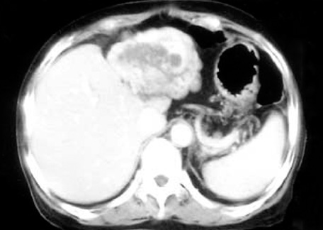
Fig. 1 Abdominal CT shows a lobulated, heterogenous contrast enhancing, soft tissue mass involving the entire left hepatic lobe (10×7 cm). Note the focal nodular enhancement around the peripheral portion of the hepatic mass in arterial phase.
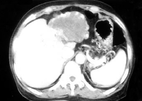
Fig. 2 Abdominal CT shows an increased extent of peripheral nodular contrast-enhancement with a persistent low density noncontrast-enhancing portion of the tumor centrally in delayed phase.
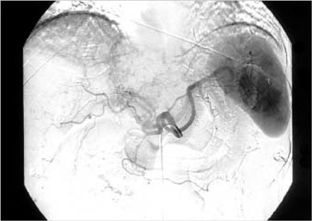
Fig. 3 Angiography shows some tumor-supplying arteries from the left hepatic artery. There is no evidence of gross invasion in the main arteries.
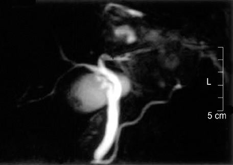
Fig. 4 MRCP shows no dilatation of intra- and extra-hepatic bile duct. The left intrahepatic duct is not visualized due to tumor replacement.
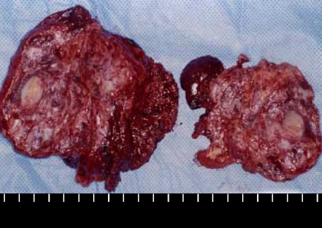
Fig. 5 The specimen shows an inhomogeneous, multinodular mass with a focal necrosis.

Fig. 6 H-E stained microscopic specimen (×40). The thrombotic material is fragmented and entrapped by the ingrowing endothelial cells. There is no soft tissue invasion, atypia, or significant necrosis.
Reference
-
1. Masson P. Hemangioendotheliome vegetant intravasculaire. Bull Soc Anat Paris. 1923. 93:517–523.2. Henschen P. L'endovasculite proliferante thrombopoietique dans la lesion vasculaire locale. Ann Anat Pathol. 1932. 9:113–121.3. Kauffman SL, Stout AP. Malignant hemangioendothelioma in infants and children. Cancer. 1961. 14:1186–1196.
Article4. Eusebi V, Fanti PA, Fedeli F, Mancini AM. Masson's intravascular vegetant hemangioendothelioma. Tumori. 1980. 66:489–498.
Article5. Kuo T, Sayers CP, Rosai J. Masson's "vegetant intravascular hemangioendothelioma:" a lesion often mistaken for angiosarcoma: study of seventeen cases located in the skin and soft tissues. Cancer. 1976. 38:1227–1236.6. Clearkin KP, Enzinger FM. Intravascular papillary endothelial hyperplasia. Arch Pathol Lab Med. 1976. 100:441–444.7. Hashimoto H, Daimaru Y, Enjoji M. Intravascular papillary endothelial hyperplasia: a clinicopathologic study of 91 cases. Am J Dermatopathol. 1983. 5:539–546.8. Amerigo J, Berry CL. Intravascular papillary endothelial hyperplasia in the skin and subcutaneous tissue. Virchows Arch A Pathol Anat Histol. 1980. 387:81–90.9. Schwartz IS, Parris A. Cutaneous intravascular papillary endothelial hyperplasia: a benign lesion that may simulate angiosarcoma. Cutis. 1982. 29:66–69. 72–74.10. Park SJ, Kim HJ, Park SH, Yeo UC, Lee ES. A case of intravascular papillary endothelial hyperplasia on upper lip. Korean J Dermatol. 2000. 38:1693–1695.11. Johraku A, Miyanaga N, Sekido N, Ikeda H, Michishita N, Saida Y, Fujiwara M, Noguchi M, Shimazui T, Akaza H. A case of intravascular papillary endothelial hyperplasia (Masson's tumor) arising from renal sinus. Jpn J Clin Oncol. 1997. 27:433–436.
Article12. Barr RJ, Graham JH, Sherwin LA. Intravascular papillary endothelial hyperplasia. A benign lesion mimicking angiosarcoma. Arch Dermatol. 1978. 114:723–726.
Article
- Full Text Links
-
- Actions
-
Cited
- CITED
-
- Close
- Share
-
- Similar articles
-
- Intravascular Papillary Endothelial Hyperplasia in Foot (A Case Report)
- Intravascular papillary endothelial hyperplasia (Masson's hemangioma) of the face
- Intravascular Papillary Endothelial Hyperplasia in Foot Adherent to a Saphenous Nerve Branch: A Case Report
- A Case of Multiple Intravascular Papillary Endothelial Hyperplasia
- Three Cases of Intravascular Papillary Endothelial Hyperplasia on the Perinasal Area
