Ileal Malignant Melanoma Presenting as a Mass with Aneurysmal Dilatation: A Case Report
- Affiliations
-
- 1Department of Surgery, College of Medicine, The Catholic University of Korea, Seoul, Korea. kimwook@hfh.cuk.ac.kr
- 2Department of Clinical Pathology, College of Medicine, The Catholic University of Korea, Seoul, Korea.
- KMID: 1733494
- DOI: http://doi.org/10.3346/jkms.2004.19.2.297
Abstract
- Malignant melanoma is the most common metastatic tumor of the gastrointestinal tract and can present with fairly common constitutional symptoms. A 36-yr-old woman was found to have a secondary malignant melanoma in the terminal ileum with profuse aneurysmal dilatation, which is not the typical presentation of the malignant melanoma in the small intestine. Radiologic studies revealed a large tumor involving the distal ileum with aneurysmal dilatations having afferent and efferent loops, which needed to be differentiated from malignant lymphoma and other gastrointestinal tumors. Exploratory laparotomy was done, and we found a huge mass with plentiful aneurysmal dilatations; much the same of the findings from the previous studies. Segmental resection with the surrounding omentum was done followed by end-to-end anastomosis between both ends of the remaining ileum. She had been free from any evidence of the local or systemic recurrence for one year after the completion of eighteen months of the subcutaneous interferon treatment; postoperatively however, the occurrence of metastatic mass at the right axilla rendered us from complete resection due to severe penetration into the vital nerves and vessels in the axilla.
MeSH Terms
Figure
-
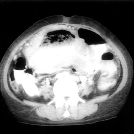
Fig. 1 Abdominal computerized tomography shows marked dilatation of the distal ileum and no obstructive lesion.

Fig. 2 Small bowel series shows submucosal tumor at distal ileum, suggesting a lymphoma with aneurysmal dilatations or gastrointestinal stromal tumor.
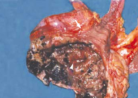
Fig. 3 There is markedly dilated lumen in the mass in connection with the lumen of afferent loop (arrowhead) and efferent loop with normal ileal mucosa (arrow).
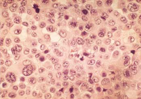
Fig. 4 The microscopic findings of the tumor cells are markedly irregular in size and shape, showing large cells with hyperchromatic nuclei, prominent nucleoli, and abundant eosinophilic cytoplasm. (H&E stain, ×400).
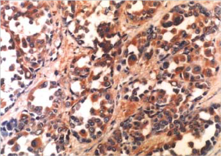
Fig. 5 Result of immunohistochemical staining with HMB-45 is positive (×200).
Cited by 1 articles
-
A Case of Primary Small Bowel Melanoma Diagnosed by Single-Balloon Enteroscopy
Jun Young Shin, In Suh Park, Byoung Wook Bang, Hyung Kil Kim, Yong Woon Shin, Kye Sook Kwon
Clin Endosc. 2017;50(4):395-399. doi: 10.5946/ce.2016.153.
Reference
-
1. Markowitz JS, Cosimi LA, Carey RW, Kang S, Padyk C, Sober AJ, Cosimi AB. Prognosis after initial recurrence of cutaneous melanoma. Arch Surg. 1991. 126:703–708.
Article2. Kim SY, Youn YK, Choe KJ. Surgical treatment of malignant melanoma. J Korean Cancer Assoc. 1990. 22:341–351.3. Das Gupta TK, Brafield RD. Metastatic melanoma: a clinicopathologic study. Cancer. 1964. 17:1323–1339.4. Klaase JM, Kroon BB. Surgery for melanoma metastatic to the gastrointestinal tract. Br J Surg. 1990. 77:60–61.
Article5. Kadivar TF, Vanek VW, Krishnan EU. Primary malignant melanoma of the small bowel: A case study. Am Surg. 1992. 58:418–422.6. Wilson BG, Anderson JR. Malignant melanoma involving the small bowel. Postgrad Med J. 1986. 62:355–357.
Article7. Patel JK, Didolkar MS, Pickren JW, Moore RH. Metastatic pattern of malignant melanoma. A study of 216 autopsy cases. Am J Surg. 1978. 135:807–810.8. Berger AC, Buell JF, Venzon D, Baker AR, Libutti SK. Management of symptomatic malignant melanoma of the gastrointestinal tract. Ann Surg Oncol. 1998. 6:155–160.
Article9. Oddson TA, Rice RP, Seigler HF, Thompson WM, Kelvin FM, Clark WM. The spectrum of small bowel melanoma. Gastrointest Radiol. 1978. 3:419–423.
Article10. Choi JY, Go YC, Kim JK, Shin SH, Cho SW, Seo KS, Kang MW, Lim YK, Yeo HS, Kim KS. A case of malignant melanoma metastasized to entire gastrointestinal tract including the esophagus. Chonnam Med J. 2000. 37:73–77.11. Sachs DL, Lowe L, Chang AE, Carson E, Johnson TM. Do primary small intestinal melanomas exist? Report of a case. J Am Acad Dermatol. 1999. 41:1042–1044.
Article12. Blecker D, Abraham S, Furth EE, Kochman ML. Melanoma in the gastrointestinal tract. Am J Gastroenterol. 1999. 94:3427–3433.
Article13. Allen AC, Spitz S. Malignant melanoma: a clinicopathological analysis of criteria for diagnosis and prognosis. Cancer. 1953. 6:1–45.14. De Matos P, Wolfe WG, Shea CR, Prieto VG, Seigler HF. Primary malignant melanoma of the esophagus. J Surg Oncol. 1997. 66:201–206.15. Norfray J, Calenoff L, Zanon B Jr. Aneurysmal lymphoma of the small intestine. Am J Roentgenol Radium Ther Nucl Med. 1973. 119:335–341.
Article16. Zornoza J, Goldstein HM. Cavitating metastases of the small intestine. Am J Roentgenol. 1977. 129:613–615.
Article17. Schuchter LM, Green R, Fraker D. Primary and metastatic diseases in malignant melanoma of the gastrointestinal tract. Curr Opin Oncol. 2000. 12:181–185.
Article18. Elsayed AM, Albahra M, Nzeako UC, Sobin LH. Malignant melanomas in the small intestine: a study of 103 patients. Am J Gastroenterol. 1996. 91:1001–1006.
- Full Text Links
-
- Actions
-
Cited
- CITED
-
- Close
- Share
-
- Similar articles
-
- Malignant Melanoma Presenting as Superior Mediastinal Mass without Extrathoracic Primary Melanoma
- Primary Pulmonary Malignant Melanoma Presenting as Bilateral Multiple Subsolid Nodules: A Case Report
- A Case of Desmoplastic Malignant Melanoma
- Primary Malignant Laryngeal Melanoma: Report of a Case with Review of Literature
- A Case of Primary Small Bowel Melanoma Diagnosed by Single-Balloon Enteroscopy
