J Vet Sci.
2009 Sep;10(3):265-267. 10.4142/jvs.2009.10.3.265.
An atypical case of respiratory actinobacillosis in a cow
- Affiliations
-
- 1Department of Veterinary Clinical Sciences, Faculty of Veterinary Medicine, Alma Mater Studiorum - University of Bologna, Italy.
- 2Department of Veterinary Public Health and Animal Pathology, Faculty of Veterinary Medicine, Alma Mater Studiorum - University of Bologna, Italy. giuliano.bettini@unibo.it
- KMID: 1726928
- DOI: http://doi.org/10.4142/jvs.2009.10.3.265
Abstract
- A not pregnant 4-year-old Jersey cow was presented with the sudden appearance of respiratory noise, nasal discharge and moderate respiratory difficulty. Upon physical examination a snoring-like noise, extended head and neck position, exaggerated abdominal effort, bilateral nasal discharge and left prescapular lymph node enlargement were noted. Sub-occlusion of the initial portion of the respiratory tract was suspected. Radiographic and endoscopic examinations revealed a pedunculate mass on the dorsal aspect of the rhinopharynx, which was removed with endoscopically assisted electrosurgery. Histologic examination revealed a chronic pyogranulomatous inflammation with eosinophilic club-like bodies surrounding small colonies of rod-shaped bacteria. Results of histochemical staining were consistent with Actinobacillus-like bacteria and a diagnosis of respiratory actinobacillosis was reached. Surgery and antibiotic therapy were resolutive, as demonstated by an endoscopic check at the second month after surgery, even without the association of the traditional iodine cure, which is regarded as the treatment of choice for actinobacillosis.
Keyword
MeSH Terms
Figure
-
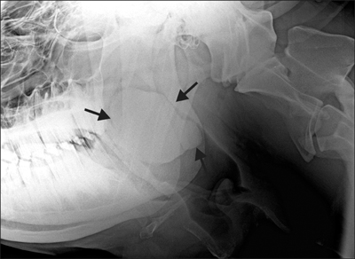
Fig. 1 Radiographic appearance of the head, latero-lateral projection. A mass is evident at the rhinopharynx level (arrows).

Fig. 2 Rhinopharyngeal actinogranuloma. The mass is composed of fibrous tissue and multiple confluent pyogranuloma. H&E stain, ×100.

Fig. 3 Actinobacillar pyogranuloma. Bacterial colonies are surrounded by eosinophilic club-like bodies, neutrophils, and large macrophages. H&E stain, ×400.
Reference
-
1. Aslani MR, Khodakaram A, Rezakhani A. An atypical case of actinobacillosis in a cow. J Vet Med A. 1995. 42:485–488.
Article2. Campbell SG, Whitlock TRH, Timoney JF, Underwood AM. An unusual epizootic of actinobacillosis in dairy heifers. J Am Vet Med Assoc. 1975. 166:604–606.3. de Kruif A, Mijten P, Haesebrouck F, Hoorens J, Devriese L. Actinobacillosis in bovine caesarean sections. Vet Rec. 1992. 131:414–415.
Article4. Gottschalk M. Actinobacillus species in animal disease: a topical subject. Vet J. 2000. 159:5–7.
Article5. Hebeler HF, Linton AH, Osborne AD. Atypical actinobacillosis in a dairy herd. Vet Rec. 1961. 73:517–521.6. Julini M, Cravero G. Actinogranulomatosi linfonodale in vitelloni da carne. Prog Vet. 1979. 34:1151–1152.7. Milne MH, Barrett DC, Mellor DJ, O'neill R, Fitzpatrick JL. Clinical recognition and treatment of bovine cutaneous actinobacillosis. Vet Rec. 2001. 148:273–274.
Article8. Radostits OM, Gay CC, Blood DC, Hinchcliff KW. Veterinary Medicine: A Textbook of the Diseases of Cattle, Sheep, Pigs, Goats and Horses. 2000. 9th ed. London: Saunders;909–944.9. Rebhun WC, King JM, Hillman RB. Atypical actinobacillosis granulomas in cattle. Cornell Vet. 1988. 78:125–130.10. Rycroft AN, Garside LH. Actinobacillus species and their role in animal disease. Vet J. 2000. 159:18–36.
Article11. Smith BP. Large Animal Internal Medicine. 2002. 3rd ed. St. Louis: Mosby;698–699.12. Swarbrick O. Atypical actinobacillosis in three cows. Br Vet J. 1967. 123:70–75.
Article
- Full Text Links
-
- Actions
-
Cited
- CITED
-
- Close
- Share
-
- Similar articles
-
- A Case of Cow's Milk Allergy with Atopic Dermatitis
- A case of occupational asthma caused by cow Hair Cross- allergenicity between cow and deer hair allergens
- A Case of Hemorrhagic Gastritis due to Cow's Milk Allergy
- Inhibitive Activity of Cow Urine and Cow Dung against Sclerotinia sclerotiorum of Cucumber
- A Case of Cow Hair-Induced Asthma in a Child Living in a Cow Raising Farm
