Polysplenia Syndrome with Congenital Agenesis of Dorsal Pancreas Presenting as Acute Pancreatitis and the Role of Endoscopic Ultrasonography in Its Diagnosis
- Affiliations
-
- 1Department of Internal Medicine, Pusan National University School of Medicine, Busan, Korea. doc0224@pusan.ac.kr
- 2Department of Radiology, Pusan National University School of Medicine, Busan, Korea.
- KMID: 1718466
- DOI: http://doi.org/10.4166/kjg.2012.60.1.47
Abstract
- A 49-year-old female was admitted to our hospital for acute pancreatitis. The abdomen CT scan incidentally showed midline liver with hepatomegaly, centrally located gallbladder, pancreas truncation, right sided small bowel, left sided large bowel, interruption of the inferior vena cava with azygos continuation, preduodenal portal vein, and multiple spleens in the left upper quadrant. In MRCP, the head of pancreas was enlarged and short main pancreatic duct without accessory duct was showed. EUS revealed enlarged ventral pancreas with a main pancreatic duct of normal caliber, absence of the accessory pancreatic duct and the dorsal pancreas. She was diagnosed as polysplenia syndrome with agenesis of dorsal pancreas. It is a rare congenital anomaly frequently associated with various visceral anomalies including multiple spleens, impaired visceral lateralization, congenital heart diseases, gastrointestinal abnormalities and azygos continuation of the inferior vena cava. We report a case of polysplenia syndrome with agenesis of dorsal pancreas presenting acute pancreatitis.
Keyword
MeSH Terms
Figure
-
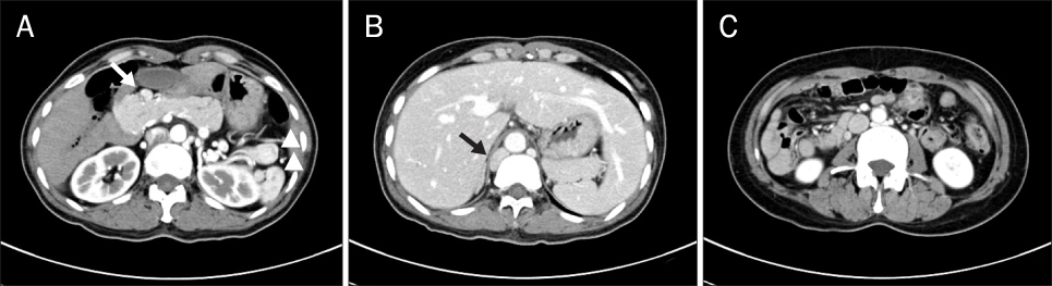
Fig. 1 Abdomen CT scans showing a midline liver, a centrally located gallbladder, multiple spleens in the left upper quadrant (arrowhead), a preduodenal portal vein (arrow), a short pancreas (A), an inferior vena cava interruption with an azygos continuation (arrow) (B), and a right-sided small bowel and left-sided large bowel (C).

Fig. 2 MRI scan (A) and MRCP (B) showing the main pancreatic duct and no accessory duct.
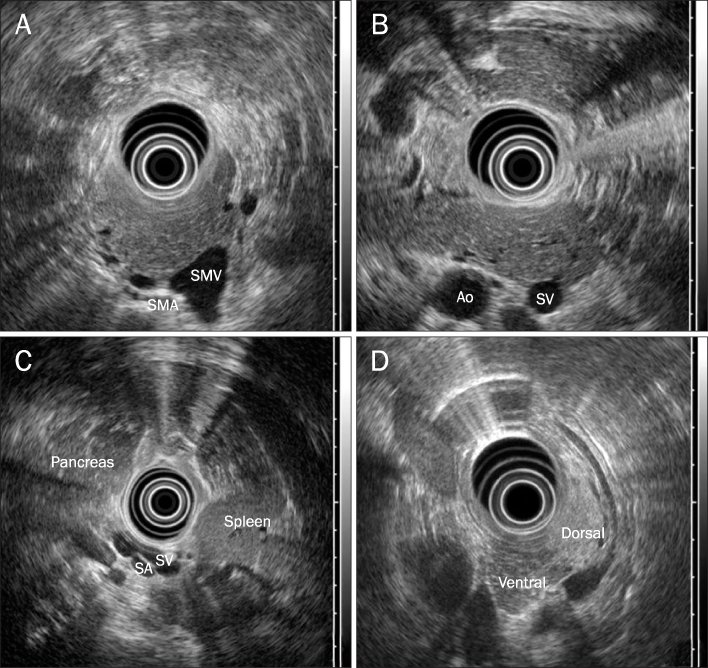
Fig. 3 EUS showing homogenous echogenicity throughout the pancreatic parenchyme (A), a hypertrophied pancreatic head, the absence of the accessory pancreatic duct (B), and splenic vessels contacting the posterior wall of the stomach (C). EUS showing different echogenicity of the ventral and dorsal pancreas in normal subject (D). SMV, superior mesenteric vein; SMA, superior mesenteric artery; Ao, aorta; SV, splenic vein; SA, splenic artery.
Reference
-
1. Fulcher AS, Turner MA. Abdominal manifestations of situs anomalies in adults. Radiographics. 2002. 22:1439–1456.2. Peoples WM, Moller JH, Edwards JE. Polysplenia: a review of 146 cases. Pediatr Cardiol. 1983. 4:129–137.3. Li CS, Tu HY, Chen RC, Yang MT, Ting CC. Polysplenia syndrome associated with preduodenal portal vein and short pancreas: Incidental findings in a case of CBD adenocarcinoma. Chin J Radiol. 2001. 26:269–274.4. Maier M, Wiesner W, Mengiardi B. Annular pancreas and agenesis of the dorsal pancreas in a patient with polysplenia syndrome. AJR Am J Roentgenol. 2007. 188:W150–W153.5. Rakesh K, Choung OW, Reddy DN. Agenesis of the dorsal pancreas (ADP) and pancreatitis - is there an association? Indian J Gastroenterol. 2006. 25:35–36.6. Palazzo L. Echoendoscopy of the pancreas. Gastroenterol Hepatol. 2002. 25:26–34.7. Tsutsumi R, Nagata Y, Enjoji A, Ohno Y, Kamito H, Kanematsu T. Situs ambiguous with gastric cancer: report of a case. Surg Today. 2007. 37:676–679.8. Hadar H, Gadoth N, Herskovitz P, Heifetz M. Short pancreas in polysplenia syndrome. Acta Radiol. 1991. 32:299–301.9. Gayer G, Apter S, Jonas T, et al. Polysplenia syndrome detected in adulthood: report of eight cases and review of the literature. Abdom Imaging. 1999. 24:178–184.10. Semb BK, Halvorsen JF. Repair of preduodenal portal vein injury occurring during biliary surgery. Acta Chir Scand. 1973. 139:312–313.11. Bobba RK, Arsura EL, Naseem M, Ashraf N. Polysplenia in an elderly male: diagnostic approaches and review of the literature. Eur J Intern Med. 2005. 16:608–609.12. Sempere L, Aparicio JR, Martínez J, Casellas JA, de Madaria E, Pérez-Mateo M. Role of endoscopic ultrasound in the diagnosis of agenesis of the dorsal pancreas. JOP. 2006. 7:411–416.13. Nishimori I, Okazaki K, Morita M, et al. Congenital hypoplasia of the dorsal pancreas: with special reference to duodenal papillary dysfunction. Am J Gastroenterol. 1990. 85:1029–1033.14. Gold RP. Agenesis and pseudo-agenesis of the dorsal pancreas. Abdom Imaging. 1993. 18:141–144.15. Suda K, Matsumoto Y, Fujii H, Miura K, Nobukawa B. Clinicopathologic differentiation of atrophy of the pancreatic body and tail aplasia. Int J Pancreatol. 1998. 24:227–235.16. Schnedl WJ, Reisinger EC, Schreiber F, Pieber TR, Lipp RW, Krejs GJ. Complete and partial agenesis of the dorsal pancreas within one family. Gastrointest Endosc. 1995. 42:485–487.17. Schnedl WJ, Piswanger-Soelkner C, Wallner SJ, et al. Agenesis of the dorsal pancreas and associated diseases. Dig Dis Sci. 2009. 54:481–487.18. Fukuoka K, Ajiki T, Yamamoto M, et al. Complete agenesis of the dorsal pancreas. J Hepatobiliary Pancreat Surg. 1999. 6:94–97.19. Wildling R, Schnedl WJ, Reisinger EC, et al. Agenesis of the dorsal pancreas in a woman with diabetes mellitus and in both of her sons. Gastroenterology. 1993. 104:1182–1186.20. Mallery S, Gupta K. Gress F, Savides T, editors. Diagnosis and staging of solid pancreatic neoplasms. Endoscopic ultrasonography. 2009. 2nd ed. West Sussex: Wiley-Blackwell;111.
- Full Text Links
-
- Actions
-
Cited
- CITED
-
- Close
- Share
-
- Similar articles
-
- Congenital Short Pancreas associated with Pancreatitis: A Case Report
- Agenesis of the Dorsal Pancreas: An autopsy case
- A Case of Partial Agenesis of Dorsal Panacreas
- A Case of Complete Agenesis of Dorsal Pancreas
- A Case of Agenesis of the Dorsal Pancreas with Concomitant Pancreatic Ductal Adenocarcinoma in an Adult Patient
