A Case of Pancreatic Neuroendocrine Tumor with Multiple Hepatic Metastasis
- Affiliations
-
- 1Department of Internal Medicine, Chonnam National University Medical School, Gwangju, Korea. choisk@chonnam.ac.kr
- KMID: 1718333
- DOI: http://doi.org/10.4166/kjg.2010.55.5.275
Abstract
- No abstract available.
MeSH Terms
-
Aged
Fluorodeoxyglucose F18/diagnostic use
Humans
Liver Neoplasms/*diagnosis/pathology/secondary
Magnetic Resonance Imaging
Male
Neuroendocrine Tumors/*diagnosis/radionuclide imaging/secondary
Pancreatic Neoplasms/*diagnosis/pathology/radionuclide imaging
Positron-Emission Tomography
Tomography, X-Ray Computed
Figure
-
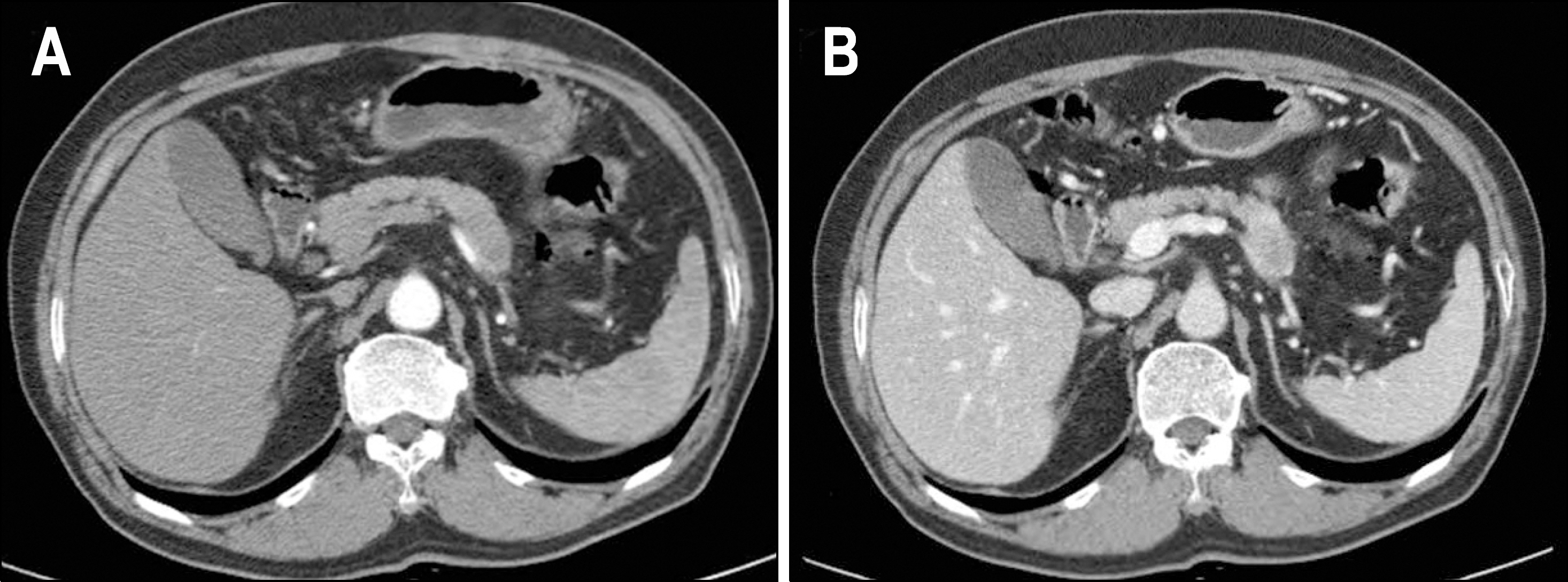
Fig. 1. (A) Early arterial phase of abdominal CT scan showed focal enlargement of the pancreas tail with peripheral enhancement. (B) Delayed venous phase of abdominal CT scan showed more strongly enhancing mass in the tail of the pancreas.

Fig. 2. (A) Abdominal MRI showed a 4 cm sized enhancing mass in the tail of the pancreas and multiple target signs in the liver. (B) MRCP showed no dilatation or obstruction of the pancreatic duct.
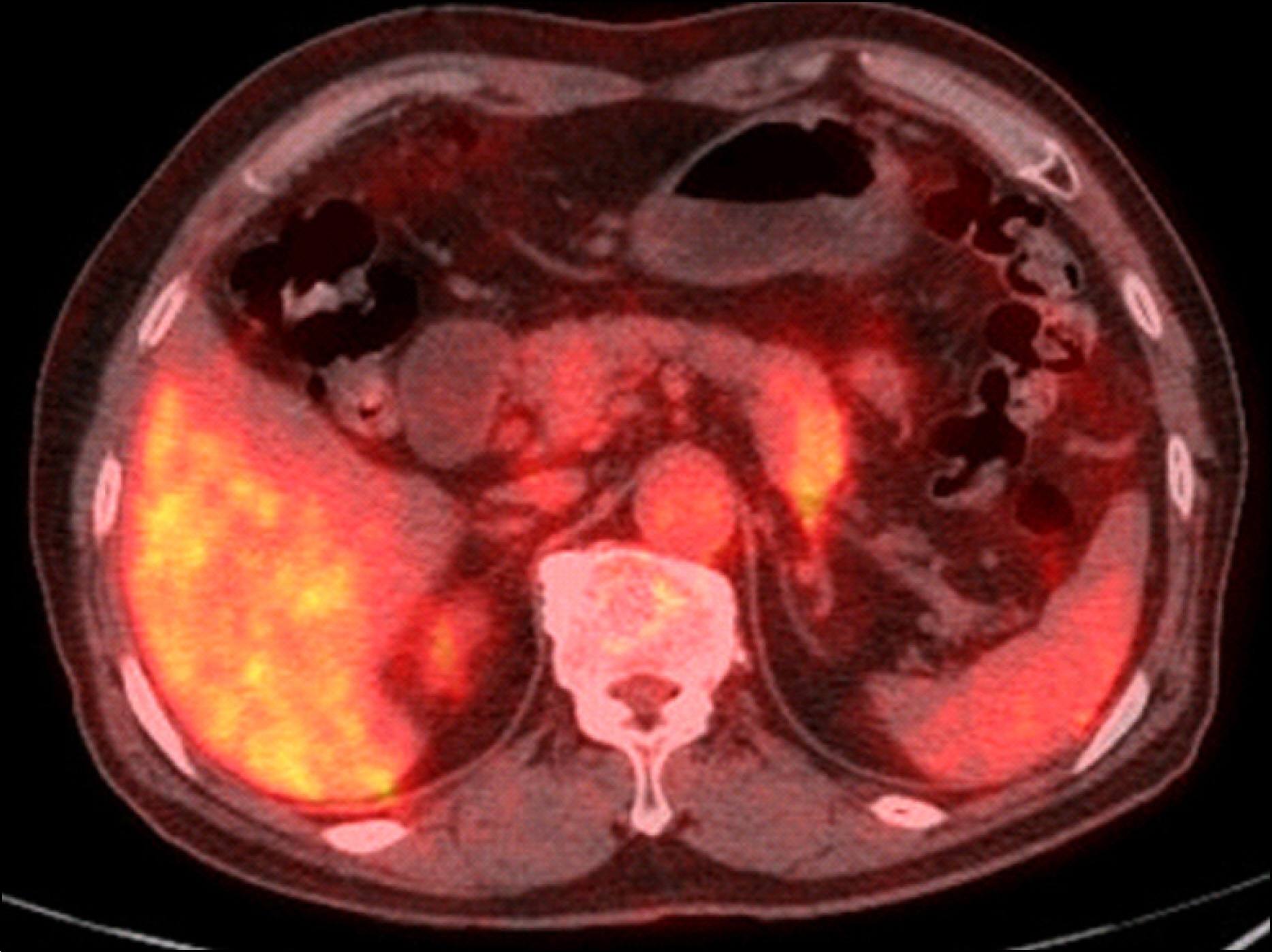
Fig. 3. FDG-PET/CT scan showed low biologic activity in the tail of the pancreas without no abnormal uptake in the liver.

Fig. 4. Liver biopsy showed (A) infiltration of small round cells with hyperchromatic nuclei (H&E stain, ×200). Tumor cell nest was strongly stained for CD 56 (B), synaptophysin (C), and cytokeratin (D, ×200).
Reference
-
1. Wick MR, Graeme-Cook FM. Pancreatic neuroendocrine neoplasms: a current summary of diagnostic, prognostic, and differential diagnostic information. Am J Clin Pathol. 2001; 115(suppl):S28–S45.2. Chetty R. An overview of practical issues in the diagnosis of gastroenteropancreatic neuroendocrine pathology. Arch Pathol Lab Med. 2008; 132:1285–1289.
Article3. Eriksson B, Arnberg H, Lindgren PG, et al. Neuroendocrine pancreatic tumors: clinical presentation, biochemical and his-topathological findings in 84 patients. J Intern Med. 1990; 228:103–113.4. Kauhanen SP, Komar G, Seppä nen MP, et al. A prospective diagnostic accuracy study of 18F-fluorodeoxyglucose positron emission tomography/computed tomography, multidetector row computed tomography, and magnetic resonance imaging in primary diagnosis and staging of pancreatic cancer. Ann Surg. 2009; 250:957–963.
Article5. Eriksson B, Ö berg K, Stridsberg M. Tumor markers in neuroendocrine tumors. Digestion. 2000; 62:33–38.
Article6. Modlin IM, Gustafsson BI, Moss SF, Pavel M, Tsolakis AV, Kidd M. Chromogranin A-biological function and clinical utility in neuro endocrine tumor disease. Ann Surg Oncol. 2010. ;[Epub ahead of print].
Article7. Simon P, Spilcke-Liss E, Wallaschofski H. Endocrine tumors of the pancreas. Endocrinol Metab Clin North Am. 2006; 35:431–447.
Article8. Delcore R, Friesen SR. Gastrointestinal neuroendocrine tumors. J Am Coll Surg. 1994; 178:187–211.9. Azimuddin K, Chamberlain RS. The surgical management of pancreatic neuroendocrine tumors. Surg Clin North Am. 2001; 81:511–525.
Article10. Basu B, Sirohi B, Corrie P. Systemic therapy for neuroendocrine tumours of gastroenteropancreatic origin. Endocr relat cancer. 2010; 17:R75–R90.
Article
- Full Text Links
-
- Actions
-
Cited
- CITED
-
- Close
- Share
-
- Similar articles
-
- Long-term Survival in Patient with Metastatic Pancreatic Neuroendocrine Tumor Treated by Variable Treatment
- Well-Differentiated Pancreatic Neuroendocrine Tumor with Solitary Hepatic Metastasis Presenting as a Benign Cystic Mass: A Case Report
- Primary Gastric Neuroendocrine Tumor with Hepatic Metastasis
- Surgical Results of Pancreatic Neuroendocrine Tumors
- Low-grade Rectal Neuroendocrine Tumor Recurring as Multiple Hepatic Metastasis after Complete Endoscopic Removal: A Case Report
