A Case of Undifferentiated (Embryonal) Liver Sarcoma Mimicking Klatskin Tumor in an Adult
- Affiliations
-
- 1Department of Internal Medicine, Kangbuk Samsung Hospital, Sungkyunkwan University School of Medicine, Seoul, Korea. hongjoo3.kim@samsung.com
- KMID: 1718299
- DOI: http://doi.org/10.4166/kjg.2010.55.2.144
Abstract
- Undifferentiated sarcoma is an uncommon primary malignant tumor of the liver typically occurring in older children. It is also referred to as malignant mesenchymoma, fibromyxosarcoma, or mesenchymal sarcoma. We experienced a case of undifferentiated sarcoma in 72-year-old male. Contrast enhanced liver CT scan revealed a 3.4 cm heterogeneously enhancing, ill-defined, and low attenuated mass in the left liver and subtle intrahepatic duct dilatation. And, in tubogram, there were segmental stenosis and occlusion from the hilum to the proximal common bile duct. We did ultrasonography guided liver biopsy. The pathologic finding revealed infiltrative growth of atypical cells with rhabdoid features. Some atypical cells showed clear cytoplasm, but no organoid pattern was identified. The stroma around atypical cells was filled with eosinophilic hyaline material. These tumor cells were positive for vimentin only, and the tumor was consistent with undifferentiated sarcoma of the liver.
MeSH Terms
-
Aged
Bile Ducts, Intrahepatic/pathology
Diagnosis, Differential
Dilatation, Pathologic
Humans
Klatskin's Tumor/diagnosis
Liver Neoplasms/*diagnosis/pathology/ultrasonography
Male
Positron-Emission Tomography
Sarcoma/*diagnosis/pathology/ultrasonography
Tomography, X-Ray Computed
Tuberculosis/drug therapy/radiography
Vimentin/metabolism
Figure
-
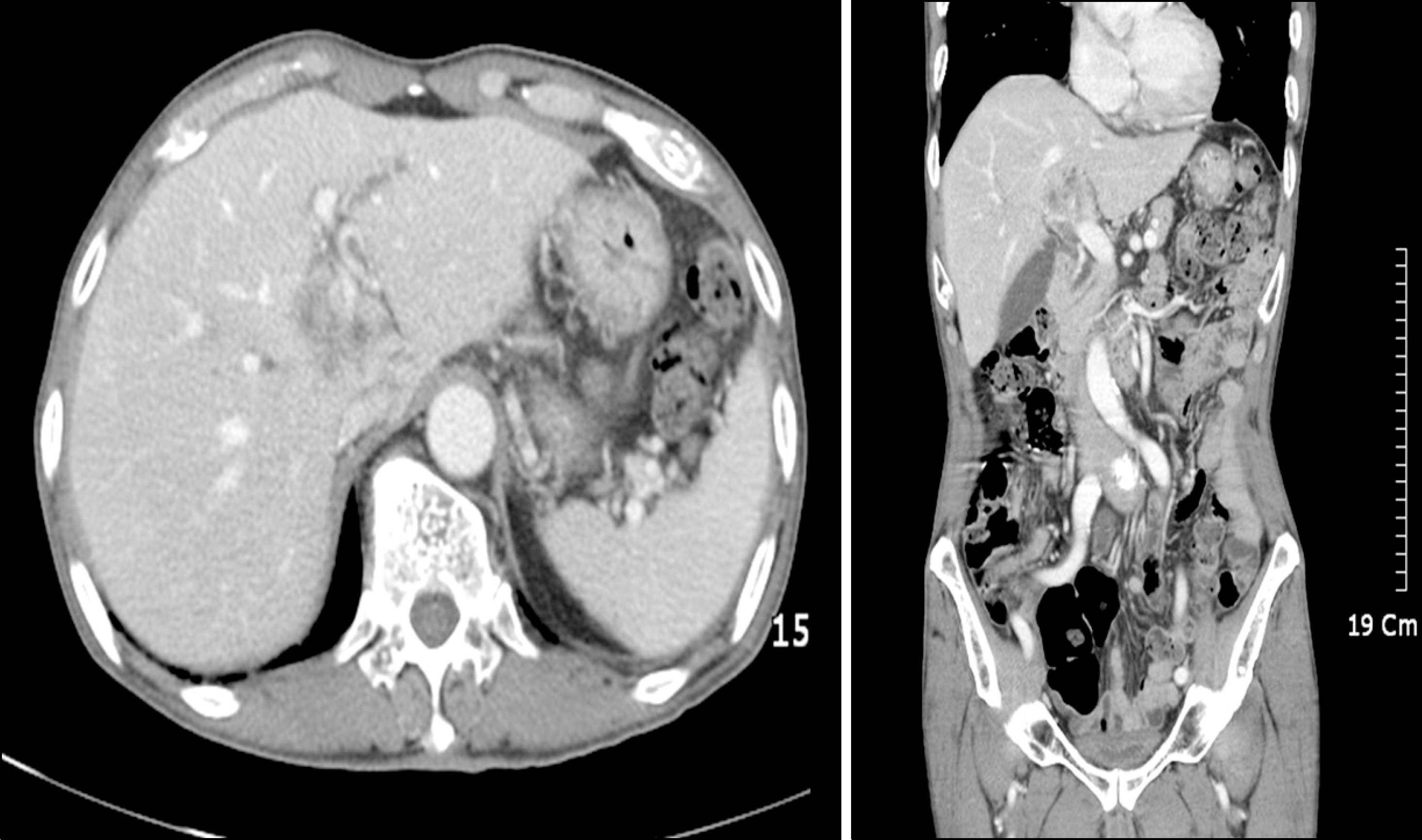
Fig. 1. Post-contrast liver CT scan. The picture showed 3.4 cm heterogenously enhancing, ill-defined, and low attenuated mass in the left lobe of the liver. There was subtle intrahepatic duct dilatation in the left liver.
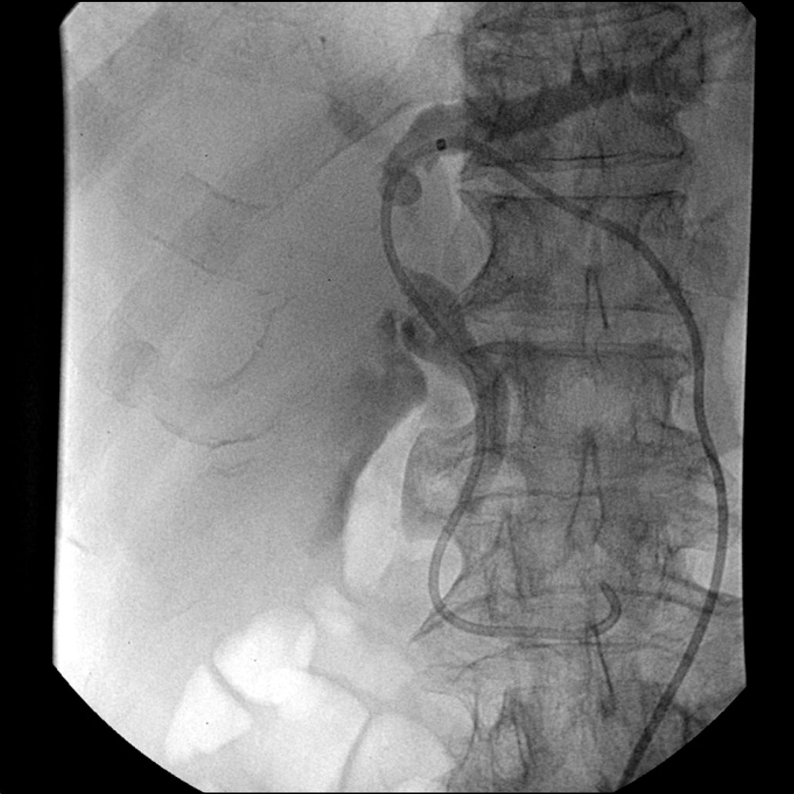
Fig. 2. Tubogram of the biliary tree. There were several segmental stenosis from the hilum to the proximal common bile duct.
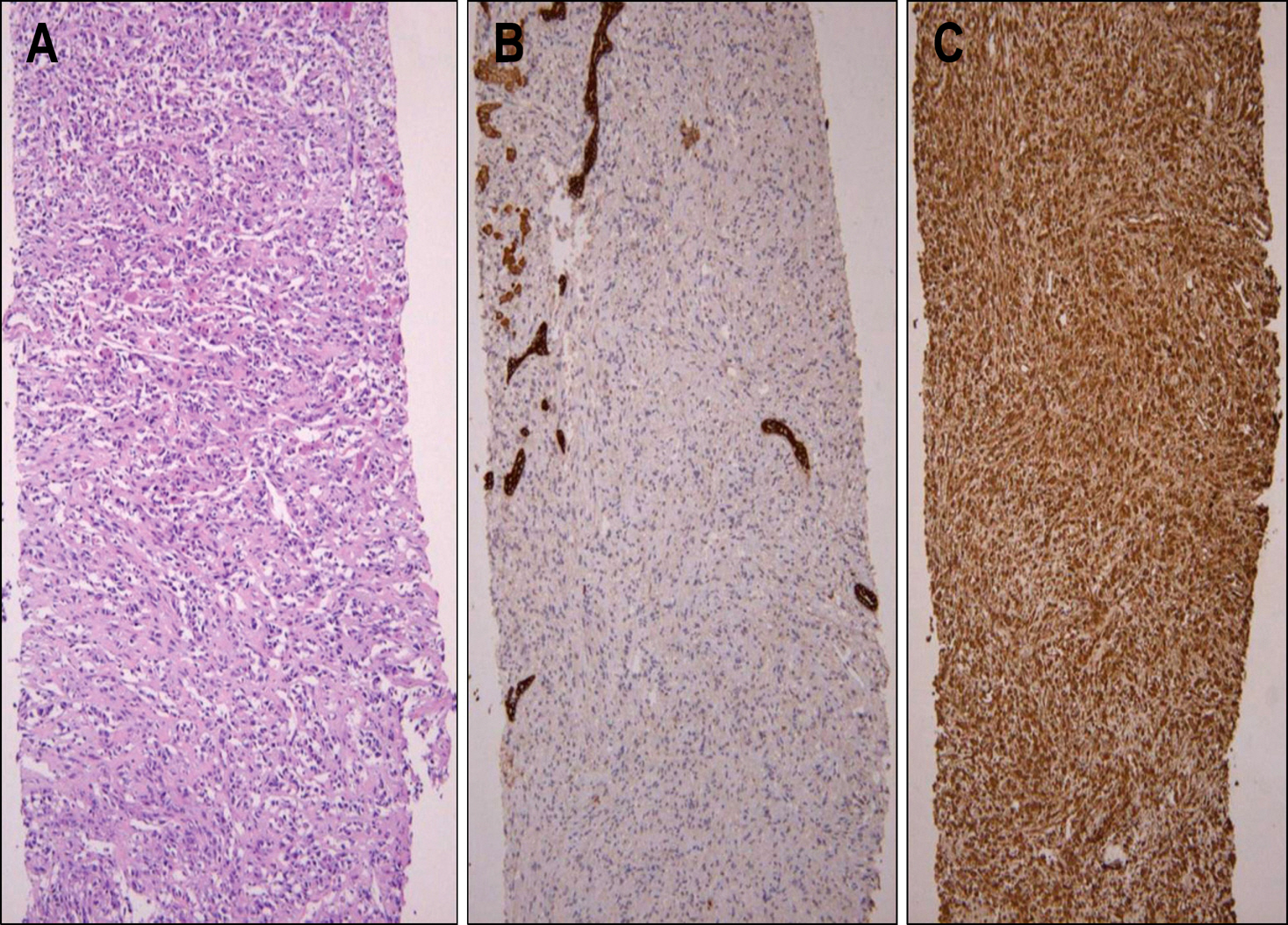
Fig. 3. Microscopic findings of the liver mass. (A) Infiltrative growth of atypical cells with rhabdoid features (H&E stain, ×200). (B) Tumor cells were negative against CK stain (CK, ×200). (C) Tumor cells are positive against vimentin stain (vimentin, ×200).

Fig. 4. Microscopic findings of the liver mass. (A) In the high power view, some atypical cells showed clear cytoplasm, but no organoid pattern was identified (H&E stain, ×400). (B) The stroma around atypical cells was filled with eosinophilic hyaline material (H&E stain, ×1,000).
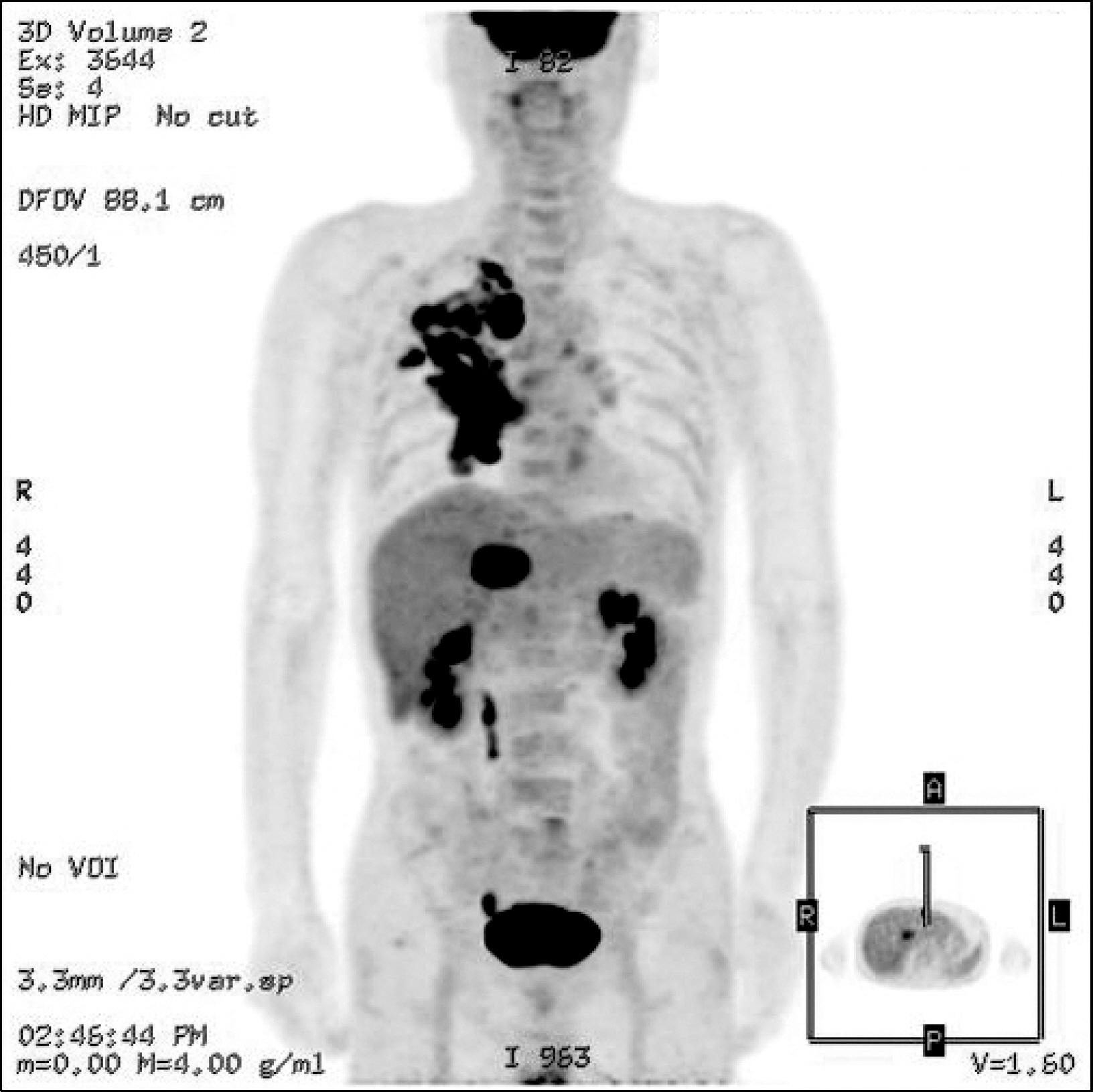
Fig. 5. Whole body PET CT. It showed a hypermetabolic mass (SUV=7.7) in central portion of the left liver segment 4. And, there was active tuberculosis with partly increased FDG uptake in the right lung. There was no metastatic lesion.
Reference
-
1. Stocker JT, Ishak KG. Undifferentiated (embryonal) sarcoma of the liver: report of 31 cases. Cancer. 1978; 42:336–348.2. Ko YK, Kim YH, Yoon YK, Yang MH, Joo HJ. Malignant mesenchymoma of the liver in the adult. K.H.M. 1986; 2:540–544.3. Chu YC, Moon YH, Kim IS. Undifferentiated sarcoma of the liver in an adult: a case report. Korean J Pathol. 1987; 21:34–39.4. Kim KH, Lee SJ, Lee G, et al. Undifferentiated sarcoma of the liver in adult: a case report and review of the literature. Korean J Hepatol. 1998; 4:283–289.5. Kim KT, Han SY, Park EH, et al. A case of the treatment in an adult with hepatic undifferentiated (embryonal) sarcoma. Korean J Hepatol. 2007; 13:96–102.6. Stanley RJ, Dehner LP, Hesker AE. Primary malignant mesenchymal tumors (mesenchymoma) of the liver in childhood. Cancer. 1973; 32:973–984.7. Chang WW, Agha FP, Morgan WS. Primary sarcoma of the liver in an adult. Cancer. 1983; 51:1510–1517.8. Miettinen M, Kahlos T. Undifferentiated (embryonal) sarcoma of the liver. Epithelial features as shown by immunohistochemical analysis and electron microscopic examination. Cancer. 1989; 64:2096–2103.
Article9. Suarez Y, De Lacy AM, Llovet JM. Images in hepatology. Intrahepatic bleeding due to undifferentiated (embryonal) hepatic sarcoma. J Hepatol. 2000; 32:361.10. Walker NI, Horn MJ, Strong RW, et al. Undifferentiated (embryonal) sarcoma of the liver. Pathologic findings and long term survival after complete surgical resection. Cancer. 1992; 69:52–59.11. Lack EE, Schloo BL, Azumi N, Travis WD, Grier HE, Kozakewich HP. Undifferentiated (embryonal) sarcoma of the liver. Clinical and pathological study of 16 cases with em-phasis on immunohistochemical features. Am J Surg Pathol. 1991; 15:1–16.12. Abramowsky CR, Cebelin M, Choudhury A, Izant RJ Jr. Undifferentiated (embryonal) sarcoma of the liver with alpha-1-antitrypsin deposits: immunohistochemical and ultrastructural studies. Cancer. 1980; 45:3108–3113.
Article13. Keating S, Taylor GP. Undifferentiated (embryonal) sarcoma of the liver: ultrastructual and immunohistochemical sim-ilarities with malignant fibrous histiocytoma. Hum Pathol. 1985; 16:693–699.14. Harris MB, Shen S, Weiner MA, et al. Treatment of primary undifferentiated sarcoma of the liver with surgery and chem-otherapy. Cancer. 1984; 54:2859–2862.
Article15. Bisgno G, Pilz T, Perilongo G, Ferrari A, Harms D, Ninfo V. Undifferentiated sarcoma of the liver in childhood; a curable disease. Cancer. 2002; 94:252–257.
- Full Text Links
-
- Actions
-
Cited
- CITED
-
- Close
- Share
-
- Similar articles
-
- Undifferentiated Embryonal Sarcoma of the Liver in an Adult: The Verification of the High Growth Rate in the Tumor
- Embryonal Sarcoma of the Liver in an Adult
- Undifferentiated embryonal sarcoma of the liver in an adult patient
- Undifferentiated Sarcoma of the Liver in Adult: A Case Report and Review of the Literature
- Undifferentiated Embryonal Sarcoma of Liver in Child
