A Case of Hyperplastic Polyp at Distal Common Bile Duct
- Affiliations
-
- 1Department of Internal Medicine, College of Medicine, The Catholic University of Korea, Seoul, Korea. isle@catholic.ac.kr
- 2Department of Pathology, College of Medicine, The Catholic University of Korea, Seoul, Korea.
- KMID: 1718290
- DOI: http://doi.org/10.4166/kjg.2010.55.2.81
Abstract
- No abstract available.
MeSH Terms
Figure
-
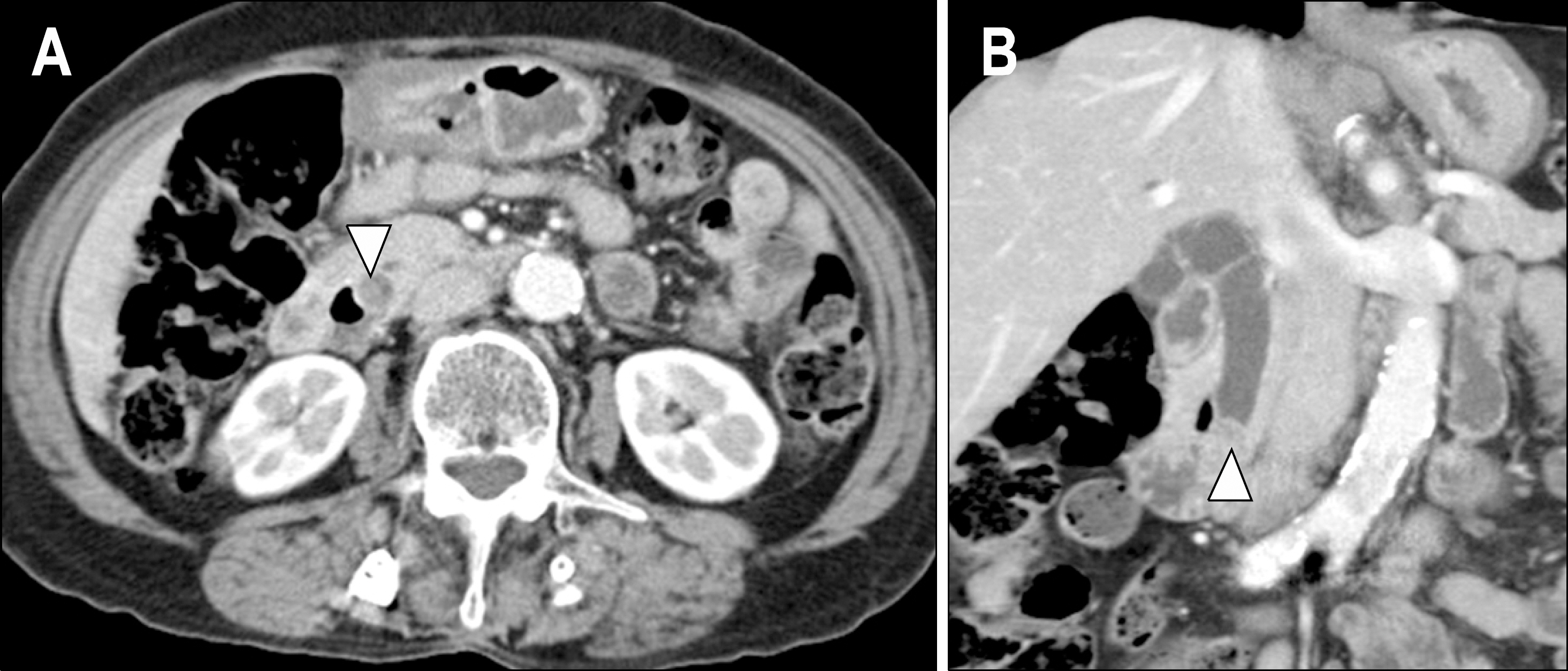
Fig. 1. Abdominal CT findings. (A) Contrast enhanced axial image. Focal enhancing polypoid lesion was noted in the ampulla of Vater or distal common bile duct. (B) Coronal image. A polypoid lesion was noted in the ampulla of Vater or distal common bile duct with dilatation of proximal common bile duct.

Fig. 2. MRI/MRCP and PET CT findings. (A) MRI image. An about 1.4 cm sized bulging contoured lesion was seen at the ampullar portion. This lesion showed enhancement after contrast infusion. (B) MRCP showed diffuse dilatation of the common bile duct and dis-tension of gallbladder with rat-tail appearing abrupt narrowing of the far-distal common bile duct. (C) PET image. Moderate FDG activity was localized in the distal portion of common bile duct.

Fig. 3. ERCP findings. (A) A large periampullary diverticulum was noted. There was no definite mass at the major ampulla. (B) An 8-mm diameter filling defect was noted at the distal common bile duct.
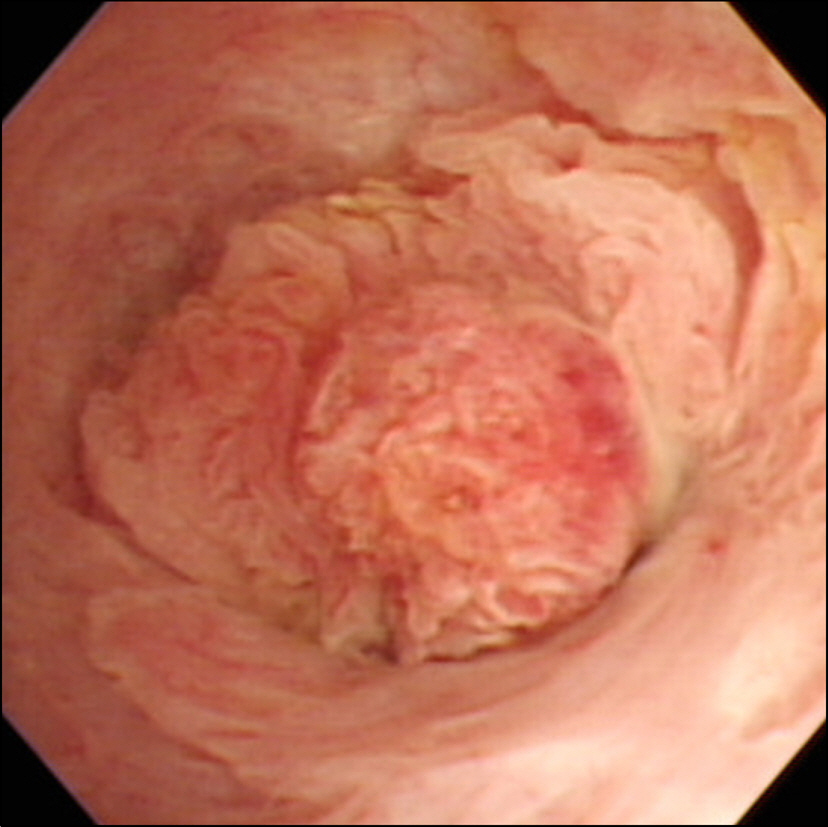
Fig. 4. Choledochoscopy showed papillary polyp-like lesion at the distal common bile duct.

Fig. 5. Pathological findings. (A) Gross examination of the specimen demonstrated a 1.2×1.0 cm polypoid lesion at distal common bile duct adjacent to major ampulla. (B) Papillary lesion was noted by H&E (×40). (C) Diffuse epithelial hyperplasia was seen by H&E (×200).
Reference
-
1. Gibson JB, Sobin LH. Histological typing of tumors of the liver, biliary tract and pancreas. Albores-Saavedra J, Henson DE, Sobin LH, editors. eds.International histological classification of tumor, No. 20. 2nd ed.Geneva: World Health Organization;1991. p. 7–8.2. Albores-Saavedra J, Henson DE. Tumors of the gallbladder and bile duct. Hartmann WH, Sobin LH, editors. eds.Atlas of tumor pathology. 2nd ed.Washington DC: Armed Forces Institute of Pathology;1984. p. 153–163.3. Inagaki M, Ishizaki A, Kino S, et al. Papillary adenoma of the distal common bile duct. J Gastroenterol. 1999; 34:535–539.
Article4. Yokohata K, Yamaguchi K, Kimura H, Tanaka M. Hyperplastic polyp of the common bile duct. Am J Gastroenterol. 1992; 87:237–239.5. Dowdy GS Jr, Olin WG Jr, Shelton EL Jr, Waldron GW. Benign tumors of the extrahepatic bile ducts. Report of three cases and review of the literature. Arch Surg. 1962; 85:503–513.6. Austin EH, Mitchell GE, Oliphant M, et al. Solitary hepatic cyst and benign bile duct polyp: a heretofore unheralded association. Surgery. 1981; 89:359–363.7. Cattell RB, Braasch JW, Kahn F. Polypoid epithelial tumors of the bile ducts. N Engl J Med. 1962; 266:57–61.
Article8. Albores-Saavedra J, Defortuna SM, Smothermon WE. Primary papillary hyperplasia of the gallbladder and cystic and common bile ducts. Hum Pathol. 1990; 21:228–231.
Article9. Ludwig J, Wahlstrom HE, Batts KP, Wiesner RH. Papillary bile duct dysplasia in primary sclerosing cholangitis. Gastroenterology. 1992; 102:2134–2138.
Article10. Fletcher ND, Wise PE, Sharp KW. Common bile duct papillary adenoma causing obstructive jaundice: case report and review of the literature. Am Surg. 2004; 70:448–452.11. Shim CS. Endoscopic imagings of the biliary tract. Chung JB, editor. ed.Diseases of the gallbladder and bile duct. 1st ed.Reston: Koonja;2008. 110-111.12. Neumann RD, LiVolsi VA, Rosenthal NS, Burrell M, Ball TJ. Adenocarcinoma in biliary papillomatosis. Gastroenterology. 1976; 70:779–782.
Article13. Ohita H, Yamaguchi Y, Yamakawa O, et al. Biliary papillomatosis with the point mutation of Kras gene arising in congenital choledochal cyst. Gastroenterology. 1993; 105:1209–1212.
Article14. Albores-Saavedra J, Henson DE, Klimstra DS. Tumors of the gallbladder, extrahepatic bile duct, and ampulla of Vater. Albores-Saavedra J, Henson DE, Klimstra DS, editors. eds.Atlas of tumor pathology. 3rd ed.Washington DC: Armed Forces Institute of Pathology;2000. p. 171–180.
- Full Text Links
-
- Actions
-
Cited
- CITED
-
- Close
- Share
-
- Similar articles
-
- A Case of Cutaneous Metastasis of Distal Common Bile Duct Carcinoma
- Clinical Significance of Hyperplastic Polyps in the Distal Colon
- A Case of Multiple Papillary Adenocarcinoma of the Extrahepatic Bile Duct : Findings of ERCP
- A Case of Villous Adenomas in the Common Bile Duct and Cystic Duct
- Computed Tomographic Findings of the Carcinoma of the Ampulla of Vater
