Diffusion-Weighted MR Imaging before and after Contrast Enhancement with Superparamagnetic Iron Oxide for Assessment of Hepatic Metastasis
- Affiliations
-
- 1Department of Radiology, Gangnam Severance Hospital, Yonsei University College of Medicine, Seoul, Korea. yjsrad97@yuhs.ac
- 2Department of Radiology, CHA Bundang Medical Center, CHA University, Seongnam, Korea.
- 3Department of Radiology, Severance Hospital, Yonsei University College of Medicine, Seoul, Korea.
- KMID: 1716885
- DOI: http://doi.org/10.3349/ymj.2012.53.4.825
Abstract
- PURPOSE
The purpose of our study was to validate diffusion-weighted MRI (DWI) before and after superparamagnetic iron oxide (SPIO) injection for assessment of hepatic metastases.
MATERIALS AND METHODS
Eighty-six hepatic metastases (size range, 0.3-4.7 cm; mean, 1.5 cm) verified pathologically or by follow-up imaging studies in 22 consecutive patients (17 men and 5 women; 44-83 years; mean age, 60 years) during a 13-month period were enrolled. Hepatic MRI, including DWI (b-factors=50, 400, 800 s/mm2) with breath-holding technique of single-shot spin-echo echo-planar imaging (TR/TE=1000/69 ms, average=2) before and after SPIO administration, were retrospectively reviewed by two independent radiologists with a 5-point scale confidence score for each hepatic lesion on pre-contrast DWI (pre-DWI), SPIO-enhanced DWI (SPIO-DWI), and SPIO-enhanced T2*-weighted imaging (SPIO-T2*wI).
RESULTS
For all lesions, SPIO-T2*wI showed significantly higher confidence score in the diagnosis of hepatic metastases than pre-contrast or SPIO-DWI regardless of the size of b-factors (p<0.05) with only one exception; using b-factor=50 s/mm2, the score of SPIO-T2*wI was still higher than SPIO-DWI but there was no statistical significance given by observer 1 (p=0.730). For the subcentimeter lesions (n=37), SPIO-T2*wI showed the highest score, and using b-factor=50 or 400 s/mm2 SPIO-DWI showed similar confidence scores to SPIO-T2*wI by both observers (p>0.05). Pre-DWI using b-factor=50 sec/mm2 was also comparable with SPIO-T2*wI by observer 1 (p=0.060).
CONCLUSION
Pre-DWI has a limited value for the assessment of hepatic metastases, however, the repetition of DWI after SPIO injection using small b-factors could complement SPIO-T2*wI, especially for subcentimeter lesions.
Keyword
MeSH Terms
Figure
-
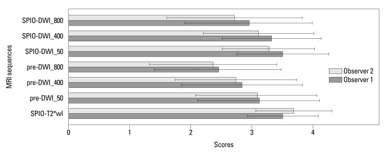
Fig. 1 Mean confidence scores with standard deviations of each MRI sequence for overall lesions. SPIO-T2*wI, SPIO-enhanced T2*wI; Pre-DWI_50, pre-contrast DWI with a b-factor of 50 s/mm2; pre-DWI_400, pre-contrast DWI with a b-factor of 400 s/mm2; pre-DWI_800, pre-contrast DWI with a b-factor of 800 s/mm2; SPIO-DWI_50, SPIO-enhanced DWI with a b-factor of 50 s/mm2; SPIO-DWI_400, SPIO-enhanced DWI with a b-factor of 400 s/mm2; SPIO-DWI_800, SPIO-enhanced DWI with a b-factor of 800 s/mm2. SPIO, superparamagnetic iron oxide; DWI, diffusion-weighted MRI; MRI, magnetic resonance imaging; T2*wI, T2* weighted imaging.
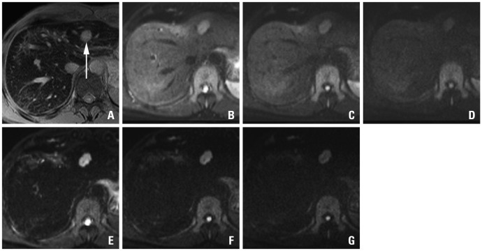
Fig. 2 A 48-year-old man with sigmoid colon cancer. SPIO-enhanced T2*-weighted image (A) shows a metastatic lesion (arrow) in the lateral segment. On the pre-contrast DWIs using b-factor (s/mm2) of 50 (B), 400 (C) and 800 (D), relative signal intensities of the lesions are gradually decreased with the increase of b-factors. On the SPIO-enhanced DWIs using b-factor (s/mm2) of 50 (E), 400 (F) and 800 (G), the lesions are more conspicuously seen, especially on the images using smaller b-factors. With an increase of b-factors, marginal blurring of the lesion looks more severe. SPIO, superparamagnetic iron oxide; DWI, diffusion-weighted MRI.
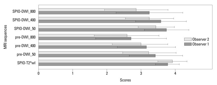
Fig. 3 Mean confidence scores with standard deviations of each MRI sequence for 1 cm or larger lesions. SPIO-T2*wI, SPIO-enhanced T2*wI; Pre-DWI_50, pre-contrast DWI with a b-factor of 50 s/mm2; pre-DWI_400, pre-contrast DWI with a b-factor of 400 s/mm2; pre-DWI_800, pre-contrast DWI with a b-factor of 800 s/mm2; SPIO-DWI_50, SPIO-enhanced DWI with a b-factor of 50 s/mm2; SPIO-DWI_400, SPIO-enhanced DWI with a b-factor of 400 s/mm2; SPIO-DWI_800, SPIO-enhanced DWI with a b-factor of 800 s/mm2. SPIO, superparamagnetic iron oxide; DWI, diffusion-weighted MRI; MRI, magnetic resonance imaging; T2*wI, T2* weighted imaging.

Fig. 4 Mean confidence scores with standard deviations of each MRI sequence for lesions smaller than 1 cm. SPIO-T2*wI, SPIO-enhanced T2*wI; Pre-DWI_50, pre-contrast DWI with a b-factor of 50 s/mm2; pre-DWI_400, pre-contrast DWI with a b-factor of 400 s/mm2; pre-DWI_800, pre-contrast DWI with a b-factor of 800 s/mm2; SPIO-DWI_50, SPIO-enhanced DWI with a b-factor of 50 s/mm2; SPIO-DWI_400, SPIO-enhanced DWI with a b-factor of 400 s/mm2; SPIO-DWI_800, SPIO-enhanced DWI with a b-factor of 800 s/mm2. SPIO, superparamagnetic iron oxide; DWI, diffusion-weighted MRI; MRI, magnetic resonance imaging; T*wI, T2* weighted imaging.
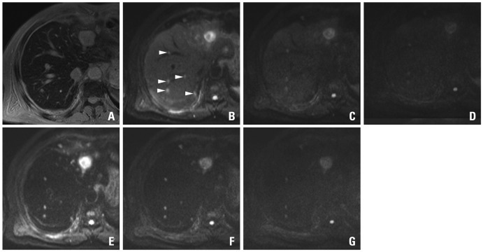
Fig. 5 A 69-year-old man with colon cancer and numerous metastases throughout the liver including the largest one in the left hemiliver. On the SPIO-enhanced T2*-weighted image (A), metastatic lesions are not well distinguished from the background liver due to small lesion size and masking effect of the intrahepatic vasculature with high signal intensity. On the pre-contrast DWIs using b-factor (s/mm2) of 50 (B), at least 5 metastases (arrowheads) in a single level of right hemiliver are well delineated from the background liver due to 'black-blood' effect. The confidence is worsened on the images using higher b-factors of 400 (C) or 800 (D) with low signal intensity and marginal blurring of the lesions. On the SPIO-enhanced DWIs using b-factor (s/mm2) of 50 (E) or 400 (F), all five lesions are conspicuously seen, compared with poor marginal definition on the image using the b-factor of 800 (G). Compared with pre-contrast DWI, SPIO-enhanced DWI using corresponding b-factors shows consistently higher lesion-to-liver contrasts. SPIO, superparamagnetic iron oxide; DWI, diffusion-weighted MRI.

Fig. 6 A 69-year-old man with multiple hepatic metastases in the left hemiliver. A small metastatatic lesion (arrowhead) on SPIO-enhanced T2*-weighted image (A) is not well distinguished from the background liver due to small size and the masking effect of the intrahepatic vasculature. On the pre-contrast DWIs using b-factor (s/mm2) of 50 (B), 400 (C) and 800 (D), there is severe motion artifact and no focal lesion is defined. On the SPIO-enhanced DWIs using b-factor (s/mm2) of 50 (E), 400 (F) and 800 (G), the lesion (arrowhead) is well delineated due to the increased lesion-to-liver contrast in spite of the motion artifact. SPIO, superparamagnetic iron oxide; DWI, diffusion-weighted MRI.
Reference
-
1. Ward J. New MR techniques for the detection of liver metastases. Cancer Imaging. 2006; 6:33–42. PMID: 16766267.
Article2. Penna C, Nordlinger B. Colorectal metastasis (liver and lung). Surg Clin North Am. 2002; 82:1075–1090. PMID: 12507210.
Article3. Robinson PJ. Imaging liver metastases: current limitations and future prospects. Br J Radiol. 2000; 73:234–241. PMID: 10817037.
Article4. Oner AY, Celik H, Oktar SO, Tali T. Single breath-hold diffusion-weighted MRI of the liver with parallel imaging: initial experience. Clin Radiol. 2006; 61:959–965. PMID: 17018309.
Article5. Erturk SM, Ichikawa T, Sano K, Motosugi U, Sou H, Araki T. Diffusion-weighted magnetic resonance imaging for characterization of focal liver masses: impact of parallel imaging (SENSE) and b value. J Comput Assist Tomogr. 2008; 32:865–871. PMID: 19204445.6. Parikh T, Drew SJ, Lee VS, Wong S, Hecht EM, Babb JS, et al. Focal liver lesion detection and characterization with diffusion-weighted MR imaging: comparison with standard breath-hold T2-weighted imaging. Radiology. 2008; 246:812–822. PMID: 18223123.
Article7. Nasu K, Kuroki Y, Nawano S, Kuroki S, Tsukamoto T, Yamamoto S, et al. Hepatic metastases: diffusion-weighted sensitivity-encoding versus SPIO-enhanced MR imaging. Radiology. 2006; 239:122–130. PMID: 16493012.
Article8. Müller MF, Prasad PV, Siewert B, Edelman RR. [The in-vivo diffusion measurements of the liver, kidneys, spleen and m. erector with an echo-planar imaging system in normal subjects]. Rofo. 1994; 161:233–236. PMID: 7919249.9. Ichikawa T, Haradome H, Hachiya J, Nitatori T, Araki T. Diffusion-weighted MR imaging with a single-shot echoplanar sequence: detection and characterization of focal hepatic lesions. AJR Am J Roentgenol. 1998; 170:397–402. PMID: 9456953.
Article10. Naganawa S, Sato C, Nakamura T, Kumada H, Ishigaki T, Miura S, et al. Diffusion-weighted images of the liver: comparison of tumor detection before and after contrast enhancement with superparamagnetic iron oxide. J Magn Reson Imaging. 2005; 21:836–840. PMID: 15906340.
Article11. Senéterre E, Taourel P, Bouvier Y, Pradel J, Van Beers B, Daures JP, et al. Detection of hepatic metastases: ferumoxides-enhanced MR imaging versus unenhanced MR imaging and CT during arterial portography. Radiology. 1996; 200:785–792. PMID: 8756932.
Article12. Vogl TJ, Schwarz W, Blume S, Pietsch M, Shamsi K, Franz M, et al. Preoperative evaluation of malignant liver tumors: comparison of unenhanced and SPIO (Resovist)-enhanced MR imaging with biphasic CTAP and intraoperative US. Eur Radiol. 2003; 13:262–272. PMID: 12598989.
Article13. Strotzer M, Gmeinwieser J, Schmidt J, Fellner C, Seitz J, Albrich H, et al. Diagnosis of liver metastases from colorectal adenocarcinoma. Comparison of spiral-CTAP combined with intravenous contrast-enhanced spiral-CT and SPIO-enhanced MR combined with plain MR imaging. Acta Radiol. 1997; 38:986–992. PMID: 9394654.14. Taouli B, Koh DM. Diffusion-weighted MR imaging of the liver. Radiology. 2010; 254:47–66. PMID: 20032142.
Article15. Hussain SM, De Becker J, Hop WC, Dwarkasing S, Wielopolski PA. Can a single-shot black-blood T2-weighted spin-echo echo-planar imaging sequence with sensitivity encoding replace the respiratory-triggered turbo spin-echo sequence for the liver? An optimization and feasibility study. J Magn Reson Imaging. 2005; 21:219–229. PMID: 15723376.
Article16. Coenegrachts K, Matos C, ter Beek L, Metens T, Haspeslagh M, Bipat S, et al. Focal liver lesion detection and characterization: comparison of non-contrast enhanced and SPIO-enhanced diffusion-weighted single-shot spin echo echo planar and turbo spin echo T2-weighted imaging. Eur J Radiol. 2009; 72:432–439. PMID: 18849130.
Article17. Kiryu S, Watanabe M, Kabasawa H, Akahane M, Aoki S, Ohtomo K. Evaluation of super paramagnetic iron oxide-enhanced diffusion-weighted PROPELLER T2-fast spin echo magnetic resonance imaging: preliminary experience. J Comput Assist Tomogr. 2006; 30:197–200. PMID: 16628031.18. Liang L, Korogi Y, Sugahara T, Shigematsu Y, Okuda T, Ikushima I, et al. Detection of intracranial hemorrhage with susceptibility-weighted MR sequences. AJNR Am J Neuroradiol. 1999; 20:1527–1534. PMID: 10512241.
- Full Text Links
-
- Actions
-
Cited
- CITED
-
- Close
- Share
-
- Similar articles
-
- Superparamagnetic Iron Oxide Enhanced MR Imaging: Influence of Hepatic Dysfunction in Cirrhotic Patients
- Cancer -Targeted MR Molecular Imaging
- Hemangioma and Hepatocellular Carcinoma: Distinction with Superparamagnetic Iron Oxide-Enhanced MR Imaging
- Intrahepatic Extramedullary Hematopoiesis Mimicking a Hypervascular Hepatic Neoplasm on Dynamic- and SPIO-Enhanced MRI
- Pharmacokinetic Modeling of Phagocytic Activity of the Liver Using Superparamagnetic Iron Oxide Nanoparticles in Dynamic MR Imaging
