Radiologic and Clinical Features of Idiopathic Granulomatous Lobular Mastitis Mimicking Advanced Breast Cancer
- Affiliations
-
- 1Department of Diagnostic Radiology, Yonsei University College of Medicine, Yongdong Severance Hospital, Seoul, Korea. kbrrdoh@yumc.yonsei.ac.kr
- 2Department of Pathology, Yonsei University College of Medicine, Yongdong Severance Hospital, Seoul, Korea.
- 3Research Institute of Radiological Science, Yonsei University, Seoul, Korea.
- 4Department of Diagnostic Radiology, Yonsei University, Wonju College of Medicine, Wonju, Korea.
- 5Department of Diagnostic Radiology, Gachon Medical School, Ghil Medical Center, Inchon, Korea.
- KMID: 1715876
- DOI: http://doi.org/10.3349/ymj.2006.47.1.78
Abstract
- Idiopathic granulomatous lobular mastitis (IGLM), also known as idiopathic granulomatous mastitis, is a rare chronic inflammatory lesion of the breast that can clinically and radiographically mimic breast carcinoma. The aim of this study was to describe the radiological imaging and clinical features of IGLM in order to better differentiate this disorder from breast cancer. We performed a retrospective analysis of the clinical and radiographic features of 11 women with a total of 12 IGLM lesions. The ages of these women ranged between 29 and 42 years, with a mean age of 34.8 years. Ten patients were examined by both mammography and sonography and one by sonography alone. The sites that were the most frequently involved were the peripheral (6/12), diffuse, (3/12), and subareolar (3/12) regions of the breast. The patient mammograms showed irregular ill-defined masses (7/11), diffuse increased densities (3/11), and one oval obscured mass. In addition, patient sonograms showed irregular tubular lesions (7/12) or lobulated masses with minimal parenchymal distortion (2/12), parenchymal distortion without definite mass lesions (2/12), and one oval mass. Subcutaneous fat obliteration (12/12) and skin thickening (11/12) were also observed in these patients. Contrary to previous reports, skin changes and subareolar involvement were not rare occurrences in IGLM. In conclusion, the sonographic features of IGLM show irregular or tubular hypoechoic masses with minimal parenchymal distortion. Both clinical information and the description of radiographic features of IGLM may aid in the differentiation between IGLM and breast cancer, however histological confirmation is still required for the proper diagnosis and treatment of the disorder.
Keyword
MeSH Terms
Figure
-
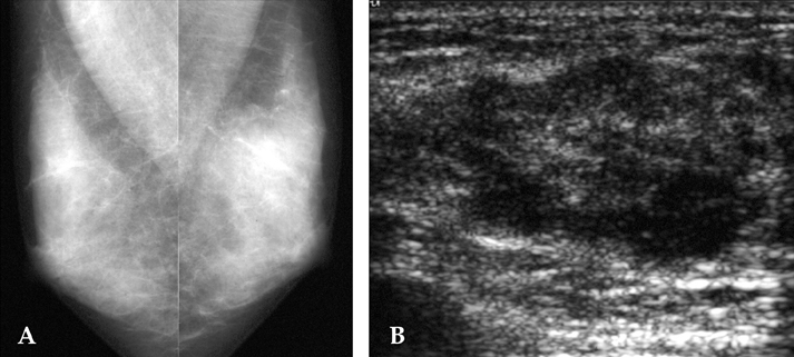
Fig. 1 Mammogram and ultrasound of a 34-year-old woman 2 months after mass detection. A. The MLO mammogram shows an irregular, indistinct high density mass in the upper left breast. B. The ultrasonography shows an ill-defined, irregular tubular, heterogeneous hypoechoic lesion.

Fig. 2 The mammogram and ultrasound of a 33-year-old woman in 2 months after the detection of a palpable mass in the left breast. A. The mammogram shows a diffuse, increased density in the left breast, but without calcification. B. The ultrasound shows parenchymal distortion without a definite mass lesion. CT scan of a 33-year-old woman in 2 months after the detection of a palpable mass in the left breast. C. The axial CT scan shows heterogeneous enhancing mass lesion with prominent skin thickening.

Fig. 3 The ultrasound and mammogram of a 34 year-old woman 2 months after the detection of a mass in the right breast. A. Ultrasonography shows a well-defined, oval, heterogeneous hypoechoic mass. B. The MR mammogram shows an abscess-like lesion with a rim enhancement in the right breast.
Cited by 1 articles
-
Medical and Surgical Treatment of Idiopathic Granulomatous Lobular Mastitis: A Benign Inflammatory Disease Mimicking Invasive Carcinoma
Gunay Gurleyik, Ali Aktekin, Fugen Aker, Hikmet Karagulle, Abdullah Saglamc
J Breast Cancer. 2012;15(1):119-123. doi: 10.4048/jbc.2012.15.1.119.
Reference
-
1. Van Ongeval C, Schraepen T, Van Steen A, Baert AL, Moerman P. Idiopathic granulomatous mastitis. Eur Radiol. 1997. 7:1010–1012.2. Jorgensen MB, Nielsen DM. Diagnosis and treatment of granulomatous mastitis. Am J Med. 1992. 93:97–101.3. Imoto S, Kitaya T, Kodama T, Hasebe T, Mukai K. Idiopathic granulomatous mastitis: case report and review of the literature. Jpn J Clin Oncol. 1997. 27:274–277.4. Han BK, Choe YH, Park JM, Moon WK, Ko YH, Yang JH, et al. Granulomatous mastitis: mammographic and sonographic appearances. AJR Am J Roentgenol. 1999. 173:317–320.5. Kessler E, Wolloch Y. Granulomatous mastitis: a lesion clinically simulating carcinoma. Am J Clin Pathol. 1972. 58:642–646.6. Going JJ, Anderson TJ, Wilkinson S, Chetty U. Granulomatous lobular mastitis. J Clin Pathol. 1987. 40:535–540.7. Donn W, Rebbeck P, Wilson C, Gilks CB. Idiopathic granulomatous mastitis. A report of three cases and review of the literature. Arch Pathol Lab Med. 1994. 118:822–825.8. Salam IM, Alhomsi MF, Daniel MF, Sim AJ. Diagnosis and treatment of granulomatous mastitis. Br J Surg. 1995. 82:214.9. Osborne BM. Granulomatous mastitis caused by histoplasma and mimicking inflammatory breast carcinoma. Hum Pathol. 1989. 20:47–52.10. Memis A, Bilgen I, Ustun EE, Ozdemir N, Erhan Y, Kapkac M. Granulomatous mastitis: imaging findings with histopathologic correlation. Clin Radiol. 2002. 57:1001–1006.11. Kocaoglu M, Somuncu I, Ors F, Bulakbasi N, Tayfun C, Ilkbahar S. Imaging findings in idiopathic granulomatous mastitis: a review with emphasis on magnetic resonance imaging. J Comput Assist Tomogr. 2004. 28:635–641.12. Rieber A, Tomczak RJ, Mergo PJ, Wenzel V, Zeitler H, Brambs HJ. MRI of the breast in the differential diagnosis of mastitis versus inflammatory carcinoma and follow-up. J Comput Assist Tomogr. 1997. 21:128–132.
