CT Evidence for Subchondral Trabecular Injury of the Femoral Head in Transient Osteoporosis of the Hip: A Case Report
- Affiliations
-
- 1Department of Orthopaedic Surgery, Inchon Medical Center Hospital, Inchon, Korea.
- 2Department of Orthopaedic Surgery, Cheju National University Hospital, Jeju, Korea.
- 3Department of Orthopaedic Surgery, Seoul National University College of Medicine, Seoul, Korea. oskim@snu.ac.kr
- 4Department of Orthopaedic Radiology, Seoul National University College of Medicine, Seoul, Korea.
- KMID: 1713858
- DOI: http://doi.org/10.3346/jkms.2010.25.1.192
Abstract
- A 28-yr-old woman presented with both hip pain that started sequentially during the peripartum period. Diagnosis of transient osteoporosis of the hip (TOH) was made based on typical findings of plain radiographs and magnetic resonance images. The subchondral trabeculae of the femoral head were evaluated on serially taken coronal multiplanar reformation computerized tomogram images. At 4 weeks after pain onset, marked decrease in the sclerotic density with irregular discontinuation was observed in the primary compression trabeculae. At 12 weeks, a focal area of irregular thickening of trabeculae was observed. At 20 weeks, sclerotic density of trabeculae recovered markedly and the focal area of irregular trabecular thickening disappeared. At 1 yr, subchondral trabeculae recovered almost completely. The evidence of subchondral trabecular injury was observed in the femoral heads of TOH.
Keyword
MeSH Terms
Figure
-
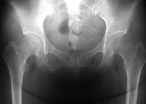
Fig. 1 A 28-yr-old woman with pain in both hips, which occurred at gestation 32 weeks in the left hip and immediately after delivery in the right. Anteroposterior radiograph obtained 1 week postpartum shows marked osteopenia in both proximal femurs.

Fig. 2 MR images taken at 2 weeks postpartum demonstrate the presence of a typical bone marrow edema pattern in both proximal femurs. T1-weighted coronal image (upper) shows decreased signal intensity area in the femoral head, neck, and intertrochanteric areas. With gadolinium enhancement (lower), the decreased intensity area was markedly enhanced.
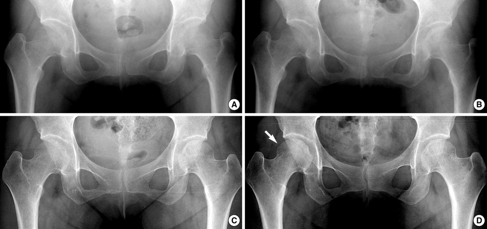
Fig. 3 Serial radiographs taken during follow-up. (A) Anteroposterior radiograph obtained at 4 weeks postpartum shows marked osteopenia in both proximal femurs. The bone density in the left side had increased and that in the right side had decreased slightly as compared with radiographs taken at 1 week postpartum (Fig. 1). (B) Anteroposterior radiograph obtained at 7 weeks postpartum shows increased osteopenia in the right femoral head. However, bone density in the left femoral head had recovered markedly. At this time, the left hip pain had almost completely disappeared. (C) Anteroposterior radiograph obtained at 11 weeks postpartum shows markedly increased bone density in both femoral heads. At this time, the right hip pain had almost completely disappeared. (D) Anteroposterior radiograph obtained at 20 weeks postpartum shows complete bone density recovery in both femoral heads. Slight dimpling (arrow) is observed in the superolateral end of the right femoral head, and this was persistent on anteroposterior radiograph taken at one year postpartum.
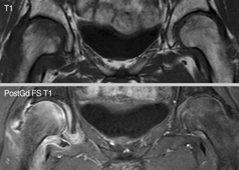
Fig. 4 On MR images taken at 7 weeks postpartum, the bone marrow edema in the left proximal femur had decreased markedly, whereas bone marrow edema persists in the right proximal femur. At this time, left hip pain had almost completely disappeared.

Fig. 5 Serial coronal 3D-CT images taken during follow-up are shown. Coronal CT image of a healthy young woman (control) shows well-oriented vertical primary compression trabeculae with normal bone density in both femoral heads. Image obtained at 4 weeks postpartum (4 w) shows reduced bone density in both proximal femurs, and a loss of thickness and continuation of vertical primary compression trabeculae. Area of irregular spotty increased bone density (arrowhead) is observed in the left femoral head immediately below the subchondral bone plate. Breakages of the subchondral bone plate (short arrows) and a sclerotic line (long arrow) obliquely crossing vertical primary compression trabeculae are observed in the right femoral head. Image taken at 11 weeks postpartum (11 w) shows a marked increase in trabecular density in both femoral heads. Area of irregular spotty bone density (arrowhead) is also observed in the right femoral head. Image taken at 20 weeks postpartum (20 w) shows almost normal appearing vertical trabeculae in both femoral heads. The irregular spotty bone density decreased. Image taken at one year postpartum (1 y) shows normalized vertical trabeculae in both femoral heads. The irregular spotty bone density has decreased further at this stage.
Reference
-
1. Guerra JJ, Steinberg ME. Distinguishing transient osteoporosis from avascular necrosis of the hip. J Bone Joint Surg Am. 1995. 77:616–624.
Article2. Song WS, Yoo JJ, Koo KH, Yoon KS, Kim YM, Kim HJ. Subchondral fatigue fracture of the femoral head in military recruits. J Bone Joint Surg Am. 2004. 86:1917–1924.
Article3. Miyanishi K, Kaminomachi S, Hara T, Maeda H, Watanabe H, Shimizu A, Torisu T. A subchondral fracture in transient osteoporosis of the hip. Skeletal Radiol. 2007. 36:677–680.
Article4. Miyanishi K, Yamamoto T, Nakashima Y, Shuto T, Jingushi S, Noguchi Y, Iwamoto Y. Subchondral changes in transient osteoporosis of the hip. Skeletal Radiol. 2001. 30:255–261.
Article5. Mitchell DG, Rao VM, Dalinka MK, Spritzer CE, Alavi A, Steinberg ME, Fallon M, Kressel HY. Femoral head avascular necrosis: correlation of MR imaging, radiographic staging, radionuclide imaging, and clinical findings. Radiology. 1987. 162:709–715.
Article6. Noorda RJ, van der Aa JP, Wuisman PI, Davis EF, Lips PT, van der Valk P. Transient osteoporosis and osteogenesis imperfecta. A case report. Clin Orthop Relat Res. 1997. 337:249–255.
Article7. Karagkevrekis CB, Ainscow DA. Transient osteoporosis of the hip associated with osteogenesis imperfecta. J Bone Joint Surg Br. 1998. 80:54–55.
Article8. Plenk H Jr, Hofmann S, Eschberger J, Gstettner M, Kramer J, Schneider W, Engel A. Histomorphology and bone morphometry of the bone marrow edema syndrome of the hip. Clin Orthop Relat Res. 1997. 334:73–84.
Article9. Todd RC, Freeman MA, Pirie CJ. Isolated trabecular fatigue fractures in the femoral head. J Bone Joint Surg Br. 1972. 54:723–728.
Article10. Yamamoto T, Schneider R, Bullough PG. Subchondral insufficiency fracture of the femoral head: histopathologic correlation with MRI. Skeletal Radiol. 2001. 30:247–254.
Article
- Full Text Links
-
- Actions
-
Cited
- CITED
-
- Close
- Share
-
- Similar articles
-
- The 1 Case Report of the Transient Osteoporosis of the Hip
- Transient osteoporosis of the hip with a femoral neck fracture during follow-up: a case report
- Current Research on Subchondral Insufficiency Fracture of the Femoral Head
- Subchondral Insufficiency Fractures of the Femoral Head
- Changes in the Microstructural and Mechanical Properties in the Medial Condyle of Human Distal Femur in Advanced Osteoarthritis
