J Korean Med Sci.
2010 Jan;25(1):152-154. 10.3346/jkms.2010.25.1.152.
Torus Hyperplasia of the Pyloric Antrum
- Affiliations
-
- 1Department of Internal Medicine, Konkuk University School of Medicine, Seoul, Korea. sunyoung@kuh.ac.kr
- 2Department of Pathology, Konkuk University School of Medicine, Seoul, Korea.
- 3Department of Surgery, Konkuk University School of Medicine, Seoul, Korea.
- KMID: 1713848
- DOI: http://doi.org/10.3346/jkms.2010.25.1.152
Abstract
- Primary or idiopathic hypertrophy of the pyloric muscle in adult, so called torus hyperplasia, is an infrequent but an established entity. It is caused by a circular muscle hypertrophy affecting the lesser curvature near the pylorus. Since most of the lesions are difficult to differentiate from tumor, distal gastrectomy is usually preformed to rule out most causes of pyloric lesions including neoplastic ones through a pathological study. A 56-yr-old man with a family history of gastric cancer presented with abdominal discomfort of 1 month duration. Upper gastrointestinal endoscopy showed a 1.0 cm sized irregular submucosal lesion proximal to the pylorus to the distal antrum on the lesser curvature. On colonoscopy examination, a 1.5 cm sized protruding mass was noticed on the appendiceal orifice. Gastrectomy and cecectomy were done, and histological section revealed marked hypertrophy of the distal circular pyloric musculature and an appendiceal mucocele. To the best of our knowledge, this is the first case of torus hyperplasia with appendiceal mucocele which is found incidentally.
Keyword
MeSH Terms
Figure
-
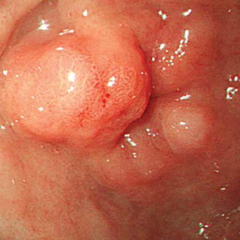
Fig. 1 Upper gastrointestinal endoscopic finding. A 10 mm sized hemispheric shaped elevated lesion with central erosion on the lesser curvature side of the distal antrum near the pylorus.
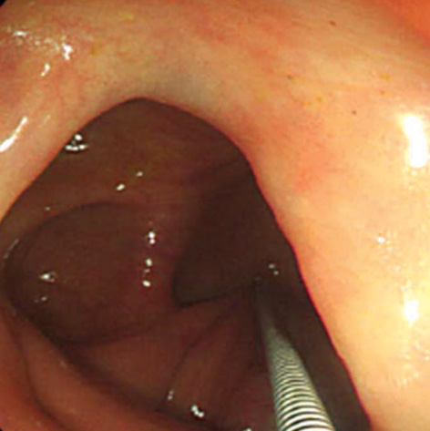
Fig. 2 Colonoscopic finding. It shows a large round mass on the cecal end with positive tent sign.
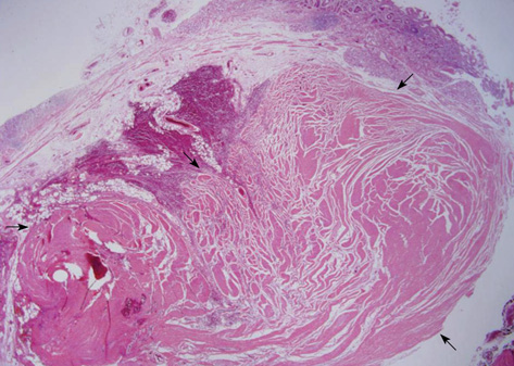
Fig. 3 Microscopic finding. In low power view, this mass-like lesion is consists of a localized area of hypertrophic circular muscle (H&E stain, ×12.5).

Fig. 4 Gross finding. Abundant mucoid material is present within the lumen of the appendix.
Reference
-
1. Aron J, Newman A, Heaton J. Torus hyperplasia of the pyloric antrum presenting as a gastric pseudotumor. Gastroenterology. 1973. 64:634–636.
Article2. Graadt van Roggen JF, M van Krieken JH. Adult hypertrophic pylroic stenosis: case report and review. J Clin Pathol. 1998. 51:479–480.3. Quigley RL, Pruitt SK, Pappas TN, Akwari O. Primary hypertrophic pyloric stenosis in the adult. Arch Surg. 1990. 125:1219–1221.
Article4. Wellmann KF, Kagan A, Fang H. Hypertorphic pyloric stenosis in adults. Gasteroenterology. 1964. 46:601–608.5. Andersen K, Gammerlgaard A, Licht E de F. Hypertrophy of the pylorus in adults. Acta Radiol. 1946. 27:552.
Article6. Ikenaga T, Honmyo U, Takano S, Murakami A, Harada K, Mizumoto S, Yoshinaka I, Hirata T, Maeda M, Kiyohara H. Primary hypertrophic pyloric stenosis in the adult. J Gastroenterol Hepatol. 1992. 7:524–526.
Article7. Dye TE, Vidals VG, Lockhart CE, Snider WR. Adult hypertrophic pyloric stenosis. Am Surg. 1979. 45:478–484.8. Hellan M, Lee T, Lerner T. Diagnosis and therapy of primary hypertrophic stenosis in adults: case report and review of literature. J Gastrointest Surg. 2006. 10:265–269.9. Franco L, Dryden N. Gastric-outlet obstruction. N Engl J Med. 2007. 356:942.
Article10. Danikas D, Peter Geis W, Ginalis E, Gorcey S, Stratoulias C. Laparascopic pyloroplasty in idiopathic hypertrophic pyloric stenosis in an adult. JSLS. 2000. 4:173–175.
- Full Text Links
-
- Actions
-
Cited
- CITED
-
- Close
- Share
-
- Similar articles
-
- Diffuse Lymphoid Hyperplsia of Gastric Antrum
- A Case of Endoscopic Temporary Stent Insertion to Treat a Pyloric Stenosis Caused by Endoscopic Submucosal Dissection for Early Gastric Cancer
- Relationship between self-reported bruxism and torus mandibularis of patients with temporomandibular disorders
- Ductal Adenocarcinoma Arising from Heterotopic Pancreas in the Stomach: A Case Report
- Dieulafoy - like Lesions of Nontraditional Locations in Gastric Antrum and Jejunum
