J Korean Med Sci.
2007 Feb;22(1):142-145. 10.3346/jkms.2007.22.1.142.
The Safety of 250 micrometer Residual Stromal Bed in Preventing Keratectasia after Laser in situ Keratomileusis (LASIK)
- Affiliations
-
- 1National Medical Center, Korea.
- 2Vision Eye Center, 581-15 Sinsa-dong, Gangnam-gu, Seoul, Korea. damholee@unitel.co.kr
- 3Department of Ophthalmology., College of Medicine, Chung-Ang University, Seoul, Korea.
- KMID: 1713254
- DOI: http://doi.org/10.3346/jkms.2007.22.1.142
Abstract
- To determine if the residual corneal stromal bed of 250 micrometer is enough to prevent iatrogenic keratectasia in laser in situ keratomileusis (LASIK), we studied 958 patients who underwent LASIK from April 2000 to October 2003 retrospectively. The estimated probabilities of the residual stromal bed, that was less than 250 micrometer, were calculated using the published flap thickness data of Moria C&B microkeratome. Then we calculated the ratio of the real incidence of keratectasia to the expected the percentage of the patients with less than 250 micrometer residual stromal bed in our study. Using the LASIK flap thickness data of Miranda, Kezirian and Nagy, the expected probabilities that the residual stroma would be less than 250 micrometer were 8.8%, 4.3% and 1.5% of the 1,916 eyes respectively, while keratectasia developed in both eyes (0.1%) of 1 patient in our study. The estimated ratio of the keratectatic eyes to eyes with less than 250 micrometer stromal bed were 1.2-6.9%. Compared to the number of eyes with residual stromal thickness less than 250 micrometer, the incidence of keratectasia was relatively low. The residual stromal bed thickness of more than 250 micrometer may possibly be safe, but further observations for long period are necessary.
MeSH Terms
Figure
-
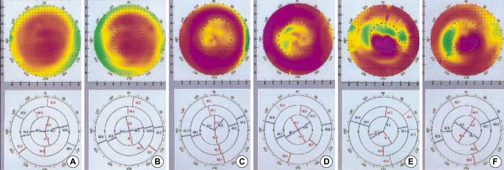
Fig. 1 Topographical changes in a patient who developed keratectasia 2 yr after LASIK. Preoperative corneal topography image. (A. Right, B. Left) Corneal topography taken 1 week after LASIK (C. Right, D. Left). Corneal topography taken 2 yr after LASIK in the right eye (E) and in the left eye (F) in the same patient. Clinically evident iatrogenic keratectasia was found 2 yr postoperatively.
Cited by 1 articles
-
Comparison of LASIK Mode Ablation and PRK Mode Ablation in LASEK Using MEL-80 Excimer Laser
Youngdon Kim, Damho Lee, Haksu Kyung
J Korean Ophthalmol Soc. 2014;55(11):1625-1630. doi: 10.3341/jkos.2014.55.11.1625.
Reference
-
1. Ou RJ, Shaw EL, Glasgow BJ. Keratectasia after laser in situ keratomileusis (LASIK): evaluation of the calculated residual stromal bed thickness. Am J Ophthalmol. 2002. 134:771–773.
Article2. Spadea L, Palmieri G, Mosca L, Fasciani R, Balestrazzi E. Iatrogenic keratectasia following laser in situ keratomileusis. J Refract Surg. 2002. 18:475–480.
Article3. Kim TG, Joo CK. 2 cases of corneal ectasia detected after LASIK. J Korean Ophthalmol Soc. 1999. 40:846–852.4. Jee DH, Kim MS. Clinical manifestation for iatrogenic keratectasia after laser in situ keratomileusis. J Korean Ophthalmol Soc. 2004. 45:920–927.5. Pallikaris IG, Kymionis GD, Astyrakakis NI. Corneal ectasia induced by laser in situ keratomileusis. J Cataract Refract Surg. 2001. 27:1796–1802.
Article6. Feltham MH, Stapleton F. Change in central corneal thickness following laser in situ keratomileusis for myopia. Clin Exp Ophthalmol. 2000. 28:185–187.
Article7. Miranda D, Smith SD, Krueger RR. Comparison of flap thickness reproducibility using microkeratomes with a second motor for advancement. Ophthalmology. 2003. 110:1931–1934.
Article8. Kezirian GM, Stonecipher KG. Comparison of the IntraLase femtosecond laser and mechanical keratomes for laser in situ keratomileusis. J Cataract Refract Surg. 2004. 30:804–811.
Article9. Nagy ZZ, Resch M, Suveges I. Ultrasound evaluation of flap thickness, ablation depth, and corneal edema after laser in situ keratomileusis. J Refract Surg. 2004. 20:279–281.
Article10. Melki SA, Azar DT. LASIK complications: Etiology, management and prevention. Surv Ophthalmol. 2001. 46:95–116.11. Seiler T, Koufala K, Richter G. Iatrogenic keratectasia after laser in situ keratomileusis. J Refract Surg. 1998. 14:312–317.
Article12. Barraquer JI. Keratomileusis for myopia and aphakia. Ophthalmology. 1981. 88:701–708.
Article13. Andreassen T, Simonsen TH, Oxlund H. Biomechanical properties of keratoconus and normal corneas. Exp Eye Res. 1980. 31:435–441.
Article14. Seiler T, Quurke AW. Iatrogenic keratectasia after LASIK in a case of forme fruste keratoconus. J Cataract Refract Surg. 1998. 24:1007–1009.
Article15. Liu KY, Lam DS. Direct measurement of microkeratome gap width by electron microscopy. J Cataract Refract Surg. 2001. 27:924–927.
Article16. Lee DH, Kwon OY, Kim JM. The comparison of corneal ablation amount by MEL-60 excimer laser and Schwind Multiscan excimer laser systems. J Korean Ophthalmol Soc. 2003. 44:1048–1053.17. Maldonado MJ, Ruiz-Oblitas L, Munuera JM, Aliseda D, Garcia-Layana A, Moreno-Montanes J. Optical coherence tomography evaluation of the corneal cap and stromal bed features after laser in situ keratomileusis for high myopia and astigmatism. Ophthalmology. 2000. 107:81–88.
Article18. Wang JC, Hufnagel TJ, Buxton DF. Bilateral keratectasia after unilateral laser in situ keratomileusis: a retrospective diagnosis of ectatic corneal disorder. J Cataract Refract Surg. 2003. 29:2015–2018.
Article19. Wirbelauer C, Pham DT. Monitoring corneal structures with slitlamp-adapted optical coherence tomography in laser in situ keratomileusis. J Cataract Refract Surg. 2004. 30:1851–1860.
Article20. Wirbelauer C, Pham DT. Intraoperative optical coherence pachymetry during laser in situ keratomileusis-first clinical experience. J Refract Surg. 2003. 19:372–377.
Article21. Casebeer JC, Slade SG, Dybbs A, Mahanti RL. Intraoperative pachometry during automated lamellar keratoplasty: a preliminary report. J Refract Corneal Surg. 1994. 10:41–44.
Article22. Dayanir V, Sakarya R, Ozcura F, Kir E, Aktunc T, Ozkan BS, Okyay P. Effect of corneal drying on central corneal thickness. J Glaucoma. 2004. 13:6–8.
Article23. Spadea L, Fasciani R, Necozione S, Balestrazzi E. Role of the corneal epithelium in refractive changes following laser in situ keratomileusis for high myopia. J Refract Surg. 2000. 16:133–139.
Article24. Ratkay-Traub I, Ferincz IE, Juhasz T, Kurtz RM, Krueger RR. First clinical results with the femtosecond neodynium-glass laser in refractive surgery. J Refract Surg. 2003. 19:94–103.
Article
- Full Text Links
-
- Actions
-
Cited
- CITED
-
- Close
- Share
-
- Similar articles
-
- Clinical Manifestation for Iatrogenic Keratectasia after Laser in Situ Keratomileusis (LASIK)
- Corneal Endothelial Morphometric Changes after Laser in Situ Keratomileusis with Residual Thickness less than 250 micro meter
- The Effect of Suturing on the Corneal Perforation During LASIK
- Change of Posterior Corneal Surface after Laser In Situ Keratomileusis; One Year Fallow-up
- Clinical Outcome of LASIK with Different Flap Thickness
