Spontaneous Closure of Iatrogenic Coronary Artery Fistula to Left Ventricle After Septal Myectomy for Hypertrophic Obstructive Cardiomyopathy
- Affiliations
-
- 1Cardiac and Vascular Center, Samsung Medical Center, Sungkyunkwan University School of Medicine, Seoul, Korea. swpark@smc.samsung.co.kr
- KMID: 1713129
- DOI: http://doi.org/10.3346/jkms.2006.21.6.1111
Abstract
- Cases of iatrogenic coronary artery fistulas draining into the left ventricle after surgical myectomy for hypertrophic obstructive cardiomyopathy have been published as sporadic reports. However, its management scheme and prognosis are not clear because of the low incidence. A 46-yr-old woman was hospitalized for evaluation of chest pain and shortness of breath for 3 months. Transthoracic echocardiographic examination showed typical hypertrophic obstructive cardiomyopathy with a peak pressure gradient of 71 mmHg across the left ventricular outflow tract. The patient underwent surgical septal myectomy. Postoperative color Doppler imaging revealed a diastolic blood flow from the interventricular septal myocardium to the left ventricular cavity, i.e. iatrogenic coronary artery fistula to the left ventricle. Ten days later, the fistula closed spontaneously which was diagnosed by transthoracic echocardiography and confirmed by coronary angiography.
MeSH Terms
Figure
-

Fig. 1 A resting electrocardiogram shows left ventricular hypertrophy with an inverted T waves in the precordial leads.
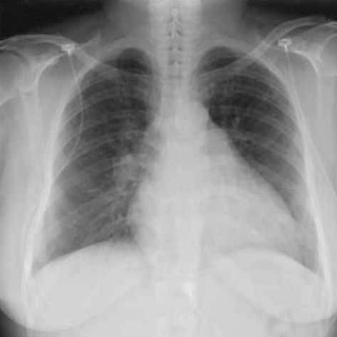
Fig. 2 Chest radiography shows moderate cardiomegaly without pulmonary edema.
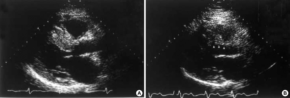
Fig. 3 Hypertrophied basal septal wall (A) was reduced in thickness after surgical septal myectomy (B). Arrowheads indicate the site of myectomy.
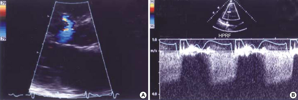
Fig. 4 In the parasternal long axis view, color Doppler examination shows the shunt flow from the septal perforators to the left ventricular cavity occurring during diastole at the myectomy site in the basal interventricular septum (A), and its peak velocity is approximately 3.0 m/sec during diastole on pulsed wave Doppler examination. It is consistent with a coronary artery to left ventricular fistula.
Reference
-
1. Hobbs RE, Millit HD, Raghavan PV, Moodie DS, Sheldon WC. Coronary artery fistulae: a 10-year review. Cleve Clin Q. 1982. 49:191–197.
Article2. Choi SH, Seo HS, Oh SJ, Hong GR, Kang SM, Lee MH, Rim SJ, Jang YS, Chung NS, Cho SY. A case of multiple coronary artery-left ventricular microfistulae demonstrated by transthoracic doppler echocardiography. Koean Circulation J. 2003. 33:338–342.
Article3. Lowe JE, Adams DH, Cummings RG, Wesly RL, Phillips HR. The natural history and recommended management of patients with traumatic coronary artery fistulas. Ann Thorac Surg. 1983. 36:295–305.
Article4. Yu R, Sharma B, Franciosa JA. Acquired coronary artery fistula to the left ventricle after acute myocardial infarction. Am J Cardiol. 1986. 58:557–558.
Article5. Sandhu JS, Uretsky BF, Zerbe TR, Goldsmith AS, Reddy PS, Kormos RL, Griffith BP, Hardesty RL. Coronary artery fistula in the heart transplant patient. A potential complication of endomyocardial biopsy. Circulation. 1989. 79:350–356.
Article6. Awasthi A, Wormer D, Heggunje PS, Obeid A. Long-term follow-up of acquired coronary artery fistula after septal myectomy for hypertrophic cardiomyopathy. J Am Soc Echocardiogr. 2002. 15:1104–1107.
Article7. Chenzbraun A, Pinto FJ, Meyer B, Stinson EB, Popp RL. Frequency of acquired coronary-cameral fistula after ventricular septal myectomy in hypertrophic cardiomyopathy. Am J Cardiol. 1993. 71:1244–1246.
Article8. Gildein HP, Kleinert S, Layangool T, Wilkinson JL. Acquired coronary artery fistula in children after ventricular septal myectomy of the right or left ventricular outflow tract. Am Heart J. 1995. 130:1124–1126.
Article9. Jebara VA, Corbi P, Dreyfus G, Acar C, Chachques JC, Rovani X, Chauvaud S, Fabiani JN, Deloche A, Carpentier A. Postoperative iatrogenic coronaro-ventricular fistula. Ann Chir. 1989. 43:643–645.10. Jebara VA, Sarkis A, Acar C, Mihaileanu S, Dervanian P, Fabiani JN, Deloche A, Laubry CP, Carpentier A. Coronary artery-left ventricle fistulas after cardiac surgery. Am Heart J. 1991. 122:1759–1762.
Article11. Marek A, Rey JL, Tribouilloy C, Jarry G, Avinee P, Lesbre JP, Quiret JC. Postoperative iatrogenic left coronaroventricular fistula. Demonstration by color two-dimensional Doppler. Arch Mal Coeur Vaiss. 1991. 84:419–423.12. Voet J, De Sutter J, De Pauw M. Diagnosis of a coronary artery fistula thirty years after myectomy for septal hypertrophy. Acta Cardiol. 2000. 55:197–198.
Article13. Bax JJ, Raphael D, Bernard X, Vanoverschelde JL. Echocardiographic detection and long-term outcome of coronary artery-left ventricle fistula after septal myectomy in hypertrophic obstructive cardiomyopathy. J Am Soc Echocardiogr. 2001. 14:308–310.
Article14. Sarkis A, Lehnert F, Diebold B, Monin JL, Cosma C, Castaldo F, Pauly-Laubry C. Left coronaro-ventricular fistula after septal myectomy. Arch Mal Coeur Vaiss. 1992. 85:457–460.15. Skimming JW, Walls JT. Congenital coronary artery fistula suggesting a "steal phenomenon" in a neonate. Pediatr Cardiol. 1993. 14:174–175.
Article16. Sapin P, Frantz E, Jain A, Nichols TC, Dehmer GJ. Coronary artery fistula: an abnormality affecting all age groups. Medicine (Baltimore). 1990. 69:101–113.
Article17. Mahoney LT, Schieken RM, Lauer RM. Spontaneous closure of a coronary artery fistula in childhood. Pediatr Cardiol. 1982. 2:311–312.
Article18. Davis JT, Allen HD, Wheller JJ, Chan DP, Cohen DM, Teske DW, Cassidy SC, Craenen JM, Kilman JW. Coronary artery fistula in the pediatric age group: a 19-year institutional experience. Ann Thorac Surg. 1994. 58:760–763.
Article19. Prewitt KC, Smolin MR, Coster TS, Vernalis MN, Bunda M, Wortham DC. Coronary artery fistula diagnosed by transesophageal echocardiography. Chest. 1994. 105:959–961.
Article20. Boxer RA, LaCorte MA, Singh S, Ishmael R, Cooper R, Stein H. Noninvasive diagnosis of congenital left coronary artery to right ventricle fistula by nuclear magnetic resonance imaging. Pediatr Cardiol. 1989. 10:45–47.
Article21. Wells RG, Litwin SB, Sty JR. Radionuclide cardioangiographic demonstration of a coronary artery fistula. Pediatr Radiol. 1986. 16:61–64.
Article22. Hofbeck M, Wild F, Singer H. Improved visualisation of a coronary artery fistula by the "laid-back" aortogram. Br Heart J. 1993. 70:272–273.
Article
- Full Text Links
-
- Actions
-
Cited
- CITED
-
- Close
- Share
-
- Similar articles
-
- Tips for Successful Septal Myectomy in Patients with Hypertrophic Cardiomyopathy
- Minimally Invasive Trans-Mitral Septal Myectomy to Treat Hypertrophic Obstructive Cardiomyopathy
- Left Ventricular Enlargement Procedure in a Patient with Diffuse-Type Hypertrophic Cardiomyopathy: A Case Report
- Early Clinical Experience of Percutaneous Transluminal Septal Myocardial Ablation and Septal Myectomy in Patients with Hypertrophic Cardiomyopathy and Severe Left Ventricular Outflow Obstruction
- Extended Septal Myectomy for Hypertrophic Obstructive Cardiomyopathy : Report of a case
