Monosegmental Hepatobiliary Fibropolycystic Disease Mimicking a Mass: Report of Three Cases
- Affiliations
-
- 1Department of Radiology, Keimyung University School of Medicine, Dongsan Medical Center, Daegu 700-712, Korea. kjh2603@dsmc.or.kr
- 2Department of Hepatobiliary Surgery, Keimyung University School of Medicine, Dongsan Medical Center, Daegu 700-712, Korea.
- 3Department of Pathology, Keimyung University School of Medicine, Dongsan Medical Center, Daegu 700-712, Korea.
- KMID: 1711477
- DOI: http://doi.org/10.3348/kjr.2014.15.1.54
Abstract
- Hepatobiliary fibropolycystic diseases are a unique group of entities involving the liver and biliary tract, which are caused by abnormal embryologic development of the ductal plates at various stages. We experienced strange hepatobiliary fibropolycystic diseases with a complex mass composed of malformed ducts and biliary cysts, which did not belong to, and were different from, previously known malformations. They were unique in imaging and histologic features. We herein report three cases of monosegmental hepatobiliary fibropolycystic disease mimicking a mass.
MeSH Terms
Figure
-
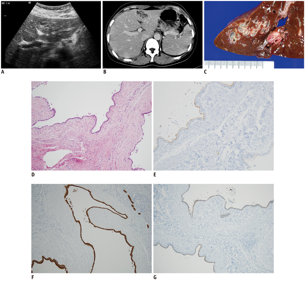
Fig. 1 Case 1. Fibropolycystic liver disease mimicking hepatic mass in 44-year-old woman. A. Transverse abdominal ultrasound shows ill-defined isoechoic solid mass with multiple internal cystic components in lateral segment of liver. B. Axial contrast-enhanced abdominal CT scan shows ill-defined cystic mass with marginal tubular components. C. Gross findings of hepatic left lobe shows aggregates of numerous small and large biliary cysts. D. Microscopic findings of mass-like monosegmental fibropolycystic disease shows numerous dilated intrahepatic bile ducts, with irregular contours, infoldings, and protrusions, surrounded by thick fibrous tissue, which were features of ductal plate malformation (hematoxylin-eosin stain, × 200). E-G. Immunohistochemical stains for CD10 (E), CK7 (F), and MUC1 (G) show positive in lining (epithelial cells) of dilated cysts, as well as those of proliferative bile ductules (× 200).
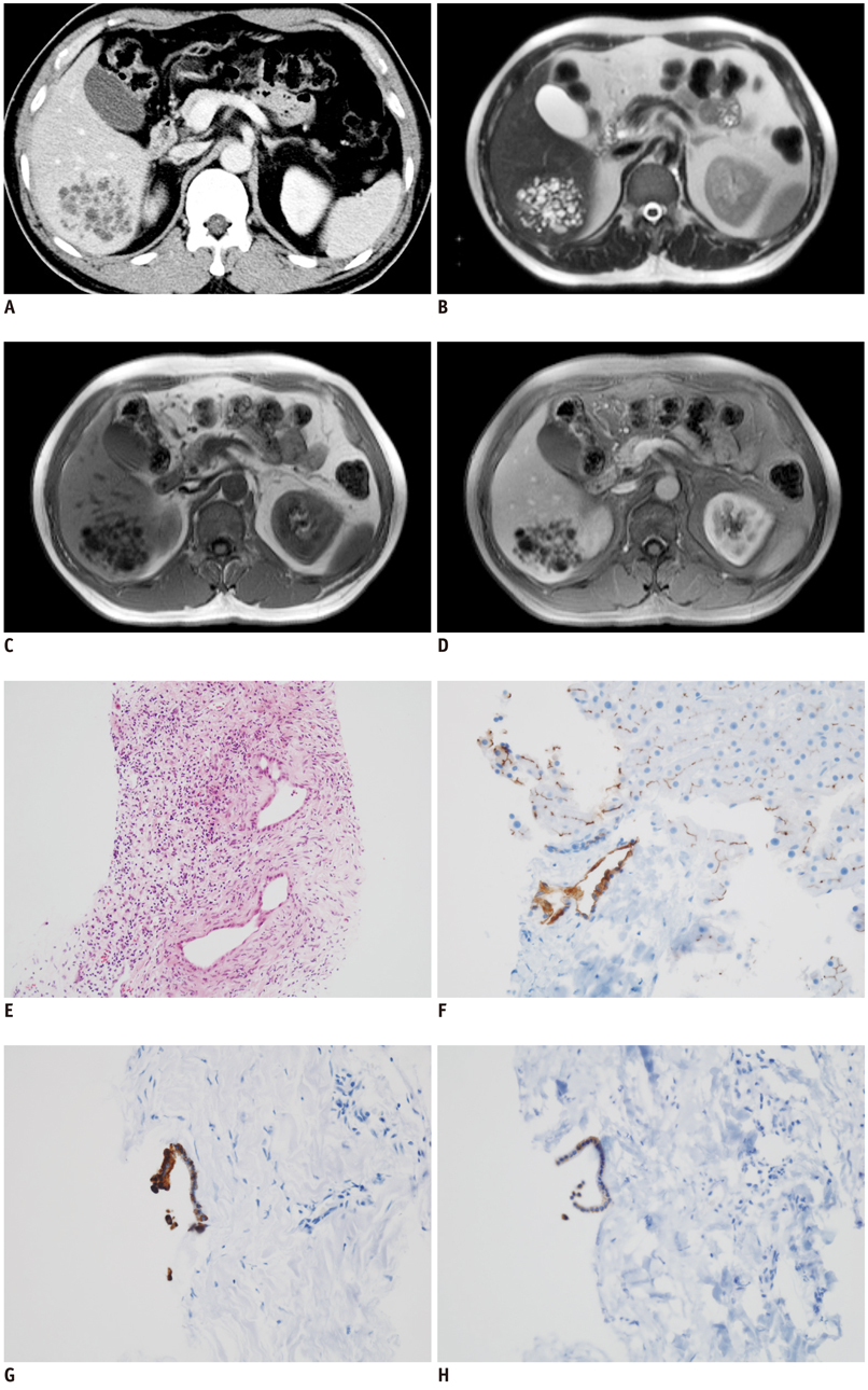
Fig. 2 Case 2. Fibropolycystic liver disease mimicking hepatic mass in 46-year-old man. A. Axial contrast-enhanced abdominal CT image shows ill-defined mass composed of numerous cysts and tubular lesions in segment VI of liver. B, C. Axial T2- and T1-weighted abdominal MR images show ill-defined mass composed of numerous cysts and tubular lesions. D. Axial contrast-enhanced abdominal MR image shows tubular and cystic lesions with no contrast enhancement. E. Microscopic finding of needle biopsy specimen shows several, irregular-shaped, dilated intrahepatic bile ducts, lined by cuboidal biliary epithelial cells. There is also ductal plate malformation and numerous inflammatory cells in background of fibrous tissue (hematoxylin-eosin stain, × 200). F-H. Immunohistochemical stains for CD10 (F), CK7 (G), MUC1 (H) shows positive in lining (epithelial cells) of dilated cysts (× 200).
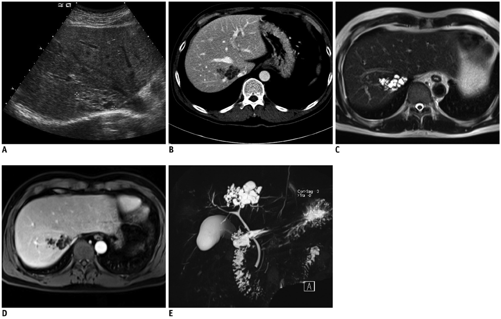
Fig. 3 Case 3. Fibropolycystic liver disease mimicking hepatic mass in 50-year-old man. A. Transverse abdominal ultrasound shows ill-defined hyperechoic solid mass with multiple internal cystic components in segment VIII of liver. B. Axial contrast-enhanced abdominal CT scan shows ill-defined cystic mass with marginal tubular components. C, D. Axial T2- and contrast-enhanced T1-weighted abdominal MR images show ill-defined mass composed of numerous cysts and tubular lesions. E. Three dimensional magnetic resonance cholangiopancreatography shows aggregates of cystic and tubular lesions.
Reference
-
1. Brancatelli G, Federle MP, Vilgrain V, Vullierme MP, Marin D, Lagalla R. Fibropolycystic liver disease: CT and MR imaging findings. Radiographics. 2005; 25:659–670.2. Levy AD, Rohrmann CA Jr, Murakata LA, Lonergan GJ. Caroli's disease: radiologic spectrum with pathologic correlation. AJR Am J Roentgenol. 2002; 179:1053–1057.3. Terada T, Moriki T. Monolobar hepatobiliary fibropolycystic disease. Pathol Oncol Res. 2011; 17:159–165.4. Terada T, Moriki T. Monolobar ductal plate malformation disease of the liver. Pathol Int. 2010; 60:407–412.5. Desmet VJ. Congenital diseases of intrahepatic bile ducts: variations on the theme "ductal plate malformation". Hepatology. 1992; 16:1069–1083.6. Summerfield JA, Nagafuchi Y, Sherlock S, Cadafalch J, Scheuer PJ. Hepatobiliary fibropolycystic diseases. A clinical and histological review of 51 patients. J Hepatol. 1986; 2:141–115.7. Veigel MC, Prescott-Focht J, Rodriguez MG, Zinati R, Shao L, Moore CA, et al. Fibropolycystic liver disease in children. Pediatr Radiol. 2009; 39:317–327. quiz 420-421.8. Lee HK, Park SJ, Yi BH, Lee AL, Moon JH, Chang YW. Imaging features of adult choledochal cysts: a pictorial review. Korean J Radiol. 2009; 10:71–80.9. Venkatanarasimha N, Thomas R, Armstrong EM, Shirley JF, Fox BM, Jackson SA. Imaging features of ductal plate malformations in adults. Clin Radiol. 2011; 66:1086–1093.10. Choi BI, Yeon KM, Kim SH, Han MC. Caroli disease: central dot sign in CT. Radiology. 1990; 174:161–163.
- Full Text Links
-
- Actions
-
Cited
- CITED
-
- Close
- Share
-
- Similar articles
-
- Fibropolycystic disease: A case report
- Two Cases of Monosegmental Pedicle Screw Fixation for Thoraco-lumbar Fracture(Three-column Injury), and a Review of the Literature
- Hilar Choledochal Cyst Mimicking Biliary Atresia on Hepatobiliary Scintigraphy: a Case Report
- Immunoglobulin G4-Related Lung Disease Mimicking Lung Cancer: Two Case Reports
- Comparison of Posterior Lumbar Interbody Fusion and Posterolateral Lumbar Fusion in Monosegmental Vacuum Phenomenon within an Intervertebral Disc
