Arachnoid Cyst in Oculomotor Cistern
- Affiliations
-
- 1Department of Radiology, Seoul St. Mary's Hospital, College of Medicine, The Catholic University of Korea, Seoul 137-701, Korea. hschoi@catholic.ac.kr
- 2Department of Neurosurgery, Seoul St. Mary's Hospital, College of Medicine, The Catholic University of Korea, Seoul 137-701, Korea.
- KMID: 1711442
- DOI: http://doi.org/10.3348/kjr.2013.14.5.829
Abstract
- Oculomotor cistern is normal anatomic structure that is like an arachnoid-lined cerebrospinal fluid-filled sleeve, containing oculomotor nerve. We report a case of arachnoid cyst in oculomotor cistern, manifesting as oculomotor nerve palsy. The oblique sagittal MRI, parallel to the oculomotor nerve, showed well-defined and enlarged subarachnoid spaces along the course of oculomotor nerve. Simple fenestration was done with immediate regression of symptom. When a disease develops in oculomotor cistern, precise evaluation with proper MRI sequence should be performed to rule out tumorous condition and prevent injury of the oculomotor nerve.
MeSH Terms
Figure
-
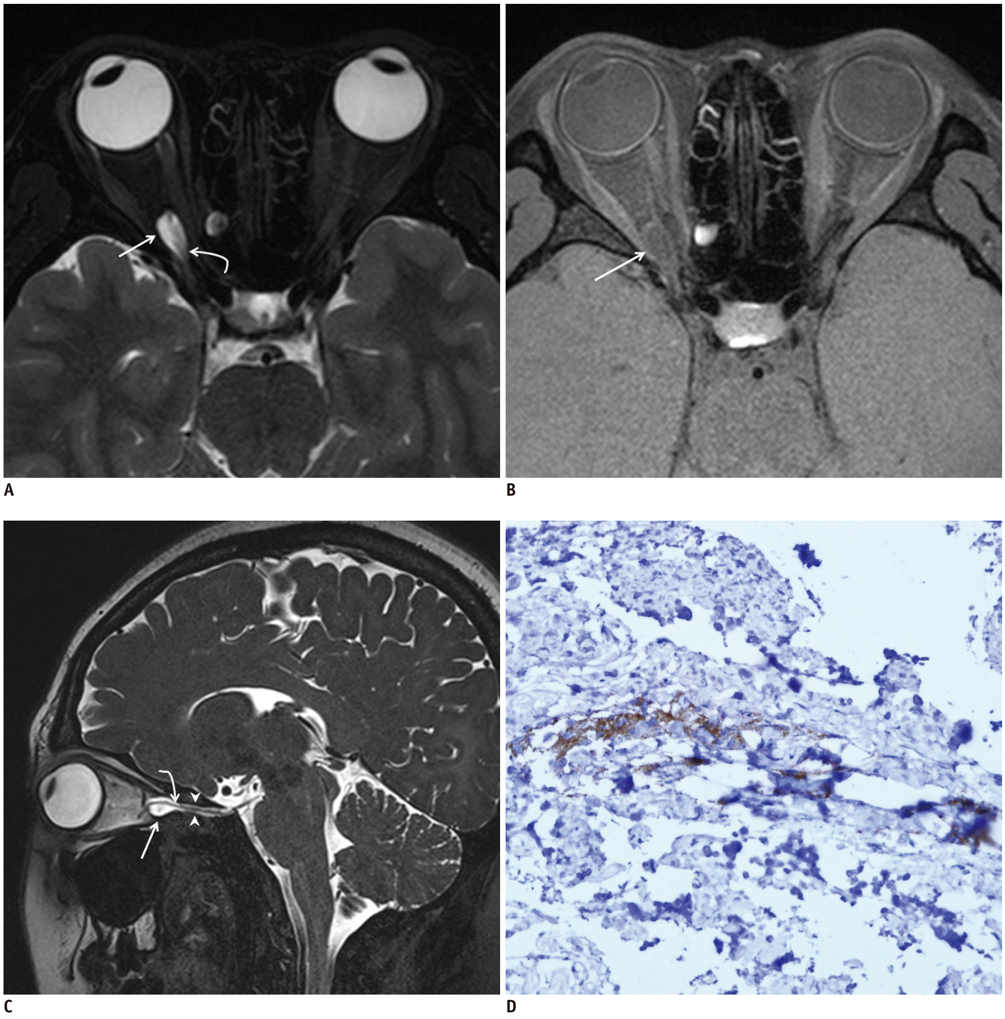
Fig. 1 Findings of arachnoid cyst in oculomotor cistern on MR images and immunohistochemistry. A. Axial MR image with heavily T2-weighted 3D turbo spin-echo sequence shows hyperintense lesion as arachnoid cyst (straight arrow) with internal oculomotor nerve (curved arrow) in right orbital apex. B. Gadolinum-enhanced T1-weighted MR image in axial plane reveals no enhancement of lesion (arrow). C. Oblique sagittal MR image with heavily T2-weighted 3D turbo spin-echo sequence reveals oculomotor nerve within lesion (curved arrow) which comes from interpeduncular cistern, passes through oculomotor cistern (between arrowheads), and continued with cystic lesion (straight arrow) in orbital apex. D. Immunohistochemical staining of surgical specimen with 400 high-power fields showed positive for epithelial membrane antigen, which is suggestive of arachnoid cyst.
Reference
-
1. Everton KL, Rassner UA, Osborn AG, Harnsberger HR. The oculomotor cistern: anatomy and high-resolution imaging. AJNR Am J Neuroradiol. 2008; 29:1344–1348.2. Martins C, Yasuda A, Campero A, Rhoton AL Jr. Microsurgical anatomy of the oculomotor cistern. Neurosurgery. 2006; 58:4 Suppl 2. ONS-220–ONS-227. discussion ONS-227-228.3. Tanriover N, Kemerdere R, Kafadar AM, Muhammedrezai S, Akar Z. Oculomotor nerve schwannoma located in the oculomotor cistern. Surg Neurol. 2007; 67:83–88. discussion 88.4. Itshayek E, Perez-Sanchez X, Cohen JE, Umansky F, Spektor S. Cavernous hemangioma of the third cranial nerve: case report. Neurosurgery. 2007; 61:E653. discussion E653.5. Yeh S, Foroozan R. Orbital apex syndrome. Curr Opin Ophthalmol. 2004; 15:490–498.6. Kim DW, Kim US. Unilateral optic nerve sheath meningocele presented with amblyopia. J Pediatr Ophthalmol Strabismus. 2011; 48 Online:e65–e66.7. Lunardi P, Farah JO, Ruggeri A, Nardacci B, Ferrante L, Puzzilli F. Surgically verified case of optic sheath nerve meningocele: case report with review of the literature. Neurosurg Rev. 1997; 20:201–205.8. Mesa-Gutiérrez JC, Quiñones SM, Ginebreda JA. Optic nerve sheath meningocele. Clin Ophthalmol. 2008; 2:661–668.9. Shanmuganathan V, Leatherbarrow B, Ansons A, Laitt R. Bilateral idopathic optic nerve sheath meningocele associated with unilateral transient cystoid macular oedema. Eye (Lond). 2002; 16:800–880.10. Khosla A, Wippold FJ 2nd. CT myelography and MR imaging of extramedullary cysts of the spinal canal in adult and pediatric patients. AJR Am J Roentgenol. 2002; 178:201–207.11. Ashker L, Weinstein JM, Dias M, Kanev P, Nguyen D, Bonsall DJ. Arachnoid cyst causing third cranial nerve palsy manifesting as isolated internal ophthalmoplegia and iris cholinergic supersensitivity. J Neuroophthalmol. 2008; 28:192–197.12. Cheng CH, Lin HL, Cho DY, Chen CC, Liu YF, Chiou SM. Intracavernous sinus arachnoid cyst with optic neuropathy. J Clin Neurosci. 2010; 17:267–269.
- Full Text Links
-
- Actions
-
Cited
- CITED
-
- Close
- Share
-
- Similar articles
-
- Neuroendoscopic Fenestration of Quadrigeminal Cistern Arachnoid Cyst Presenting with Developmental Regression
- Evaluation of the Arachnoid Cyst Treatment
- A Case of Prenatal diagnosis and Postnatal Treatment of Suprasellar Arachnoid Cyst
- A Case of Suprasellar Arachnoid Cyst
- Intracystic Hemorrhage of an Arachnoid Cyst: a Case with Prediagnostic Imaging of an Intact Cyst
