Korean J Gastroenterol.
2014 Jan;63(1):42-46. 10.4166/kjg.2014.63.1.42.
An Arteriovenous Malformation in the Jejunum Mimicking a Gastrointestinal Stromal Tumor
- Affiliations
-
- 1Department of Gastroenterology, Asan Medical Center, University of Ulsan College of Medicine, Seoul, Korea. dohoon.md@gmail.com
- 2Department of Pathology, Asan Medical Center, University of Ulsan College of Medicine, Seoul, Korea.
- KMID: 1711305
- DOI: http://doi.org/10.4166/kjg.2014.63.1.42
Abstract
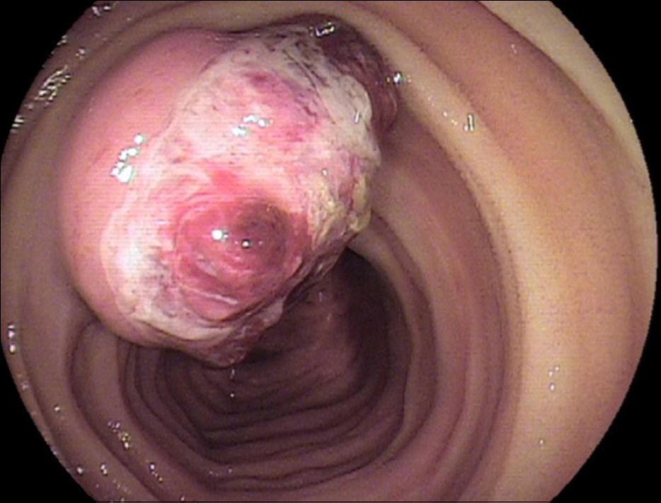
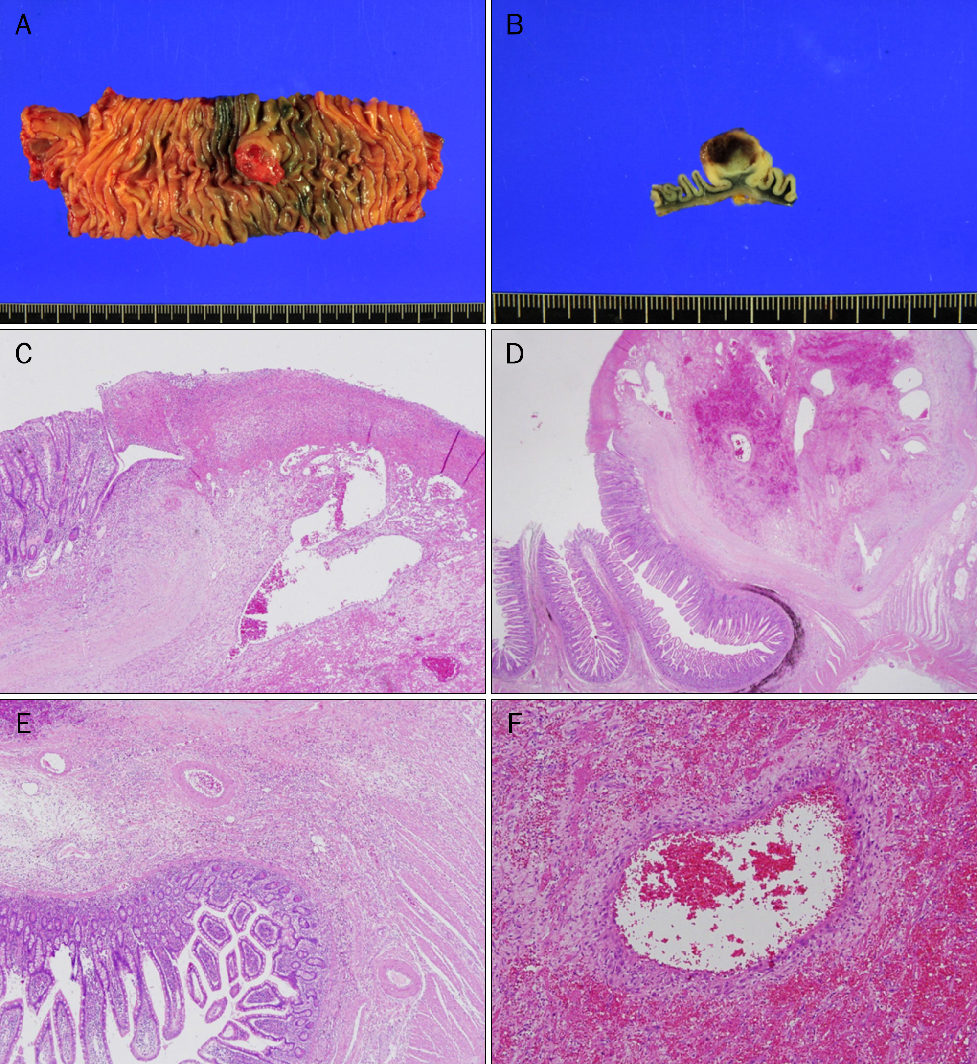
- A 51-year-old man visited the tertiary-care hospital with a 2-week history of dizziness and dyspnea on exertion. The initial hemoglobin level was 5.8 g/dL, without any history of hematochezia or melena. The esophagogastroduodenoscopy (EGD) was normal. During colonoscopic preparation, the patient experienced hematochezia and became hypotensive. On angiography, no extravasation of contrast media was observed. A CT scan with angiography showed a small high-density area in the jejunal lumen, suggesting extravasation of the contrast media. Capsule endoscopy was performed, and oozing bleeding was suspected in the proximal to mid jejunum. The patient was referred to our hospital. Repeated EGD and CT enterography did not reveal any significant bleeding. An antegrade double balloon endoscopy was performed, and an approximately 2-cm-sized submucosal tumor with ulceration and a non-bleeding exposed vessel was observed in the mid jejunum. The presumed diagnosis was jejunal gastrointestinal stromal tumor. The mass was surgically resected, and the final histopathological diagnosis was arteriovenous malformation.
MeSH Terms
Figure
Cited by 1 articles
-
Endoscopic Resection of Colonic Vascular Ectasia Mimicking as a Pedunculated Polypoid Lesion
Sang Hoon Lee, Sung Chul Park, Sung Joon Lee, Seung-Joo Nam, Seung Koo Lee
Korean J Gastroenterol. 2019;73(6):370-372. doi: 10.4166/kjg.2019.73.6.370.
Reference
-
References
1. Gordon FH, Watkinson A, Hodgson H. Vascular malformations of the gastrointestinal tract. Best Pract Res Clin Gastroenterol. 2001; 15:41–58.
Article2. Rodriguez-Jurado R, Morales SS. Polypoid arteriovenous malformation in the jejunum of a child that mimics intussusception. J Pediatr Surg. 2010; 45:E9–E12.
Article3. Yazbeck N, Mahfouz I, Majdalani M, Tawil A, Farra C, Akel S. Intestinal polypoid arteriovenous malformation: unusual presentation in a child and review of the literature. Acta Paediatr. 2011; 100:e141–e144.
Article4. Kaler SG, Westman JA, Bernes SM, et al. Gastrointestinal hemorrhage associated with gastric polyps in Menkes disease. J Pediatr. 1993; 122:93–95.
Article5. Meyer CT, Troncale FJ, Galloway S, Sheahan DG. Arteriovenous malformations of the bowel: an analysis of 22 cases and a review of the literature. Medicine (Baltimore). 1981; 60:36–48.6. Cheon JH, Song HJ, Kim JS, et al. Recurrent lower gastrointestinal bleeding from congenital arteriovenous malformation in the terminal ileum mimicking intestinal varicosis: a case report. J Korean Med Sci. 2007; 22:746–749.
Article7. Maeng L, Choi KY, Lee A, Kang CS, Kim KM. Polypoid arteriovenous malformation of colon mimicking inflammatory fibroid polyp. J Gastroenterol. 2004; 39:575–578.
Article8. Krinsky ML, Robert ME, Garcia JC, Korzenik JR, Topazian M. Polypoid vascular malformations of the small intestine. Gastrointest Endosc. 1998; 48:530–533.
Article9. Moore JD, Thompson NW, Appelman HD, Foley D. Arteriovenous malformations of the gastrointestinal tract. Arch Surg. 1976; 111:381–389.
Article10. De Palma GD, Aprea G, Rega M, et al. Polypoid vascular malformation of the small intestine. Gastrointest Endosc. 2007; 65:328–329.
Article11. Jain R, Chetty R. Dieulafoy disease of the colon. Arch Pathol Lab Med. 2009; 133:1865–1867.
Article12. Yano T, Yamamoto H, Sunada K, et al. Endoscopic classification of vascular lesions of the small intestine (with videos). Gastrointest Endosc. 2008; 67:169–172.
Article13. Leighton JA, Goldstein J, Hirota W, et al. Obscure gastrointestinal bleeding. Gastrointest Endosc. 2003; 58:650–655.
Article14. Chong AK, Chin BW, Meredith CG. Clinically significant smallbowel pathology identified by double-balloon enteroscopy but missed by capsule endoscopy. Gastrointest Endosc. 2006; 64:445–449.
Article15. Endo H, Matsuhashi N, Inamori M, et al. Tumorous arteriovenous malformation in the jejunum missed by capsule endoscopy. Gastrointest Endosc. 2008; 68:773–774.
Article16. Cui J, Huang LY, Wu CR. Small intestinal vascular malformation bleeding: diagnosis by double-balloon enteroscopy combined with abdominal contrast-enhanced CT examination. Abdom Imaging. 2012; 37:35–40.
Article17. Hansel SL, Decker GA, Shiff AD. Thirty years of overt, obscure GI bleeding solved by modern technology. Gastrointest Endosc. 2009; 70:595–597.
Article18. Nakabayashi T, Kudo M, Hirasawa T, Kuwano H. Arteriovenous malformation of the jejunum detected by arterialphase enhanced helical CT, a case report. Hepatogastroenterology. 2004; 51:1066–1068.19. Ji JS, Choi KY, Lee BI, et al. A large polypoid arteriovenous malformation of the colon treated with a detachable snare: case report and review of literature. Gastrointest Endosc. 2005; 62:172–175.
Article20. Liao Z, Gao R, Li ZS. Vascular malformation of the small intestine. Endoscopy. 2007; 39(Suppl 1):E319.
Article
- Full Text Links
- Actions
-
Cited
- CITED
-
- Close
- Share
- Similar articles
-
- A Case of Massive Bleeding from Jejunal Stromal Tumor Diagnosed by Intraoperative Enteroscopy: A Case of Jejunal Stromal Tumor Bleeding
- Synchronous Development of Gastrointestinal Stromal Tumor and Arteriovenous Malformation in the Jejunum: A Case Report
- Multiple Gastrointestinal Stromal Tumors of the Small Intestine
- Unusual Giant Arteriovenous Malformation in Jejunum: A Case Report
- Primary Gastrointestinal Stromal Tumor Accompanied with Gastrointestinal Bleeding


