Clin Orthop Surg.
2013 Sep;5(3):202-208. 10.4055/cios.2013.5.3.202.
The Prevalence of Os Acromiale in Korean Patients Visiting Shoulder Clinic
- Affiliations
-
- 1Department of Orthopaedic Surgery, Samsung Medical Center, Sungkyunkwan University School of Medicine, Seoul, Korea. shoulderyoo@gmail.com
- 2Department of Physical Medicine & Rehabilitation, Samsung Medical Center, Sungkyunkwan University School of Medicine, Seoul, Korea.
- 3Madi Hospital, Seoul, Korea.
- 4Department of Orthopaedic Surgery, Gangneung Asan Hospital, University of Ulsan College of Medicine, Gangneung, Korea. hyunil.lee7@gmail.com
- KMID: 1705546
- DOI: http://doi.org/10.4055/cios.2013.5.3.202
Abstract
- BACKGROUND
The prevalence of os acromiale has been documented to be between 1% and 15% and is known to be clinically associated with subacromial impingement or rotator cuff tear. However, the prevalence of os acromiale in Korea has not yet been determined. The purpose of this study is to evaluate the prevalence of os acromiale in Korean patients who visited shoulder clinics and to investigate the correlations with rotator cuff tear.
METHODS
We retrospectively reviewed the X-rays of patients visiting a shoulder clinic at a tertiary hospital in Korea from January 2011 to January 2012 to determine the frequency of os acromiale. X-ray findings were confirmed with magnetic resonance imaging (MRI) for patients who had these images available. MRI was also used to assess the status of the rotator cuff. The correlation between the presence of os acromiale either with gender, hand dominance or rotator cuff tear was analyzed statistically.
RESULTS
A total of 2,946 shoulders from 1,568 patients were analyzed with X-rays. Thirteen cases out of 1,568 patients had an os acromiale; and there were five and eight cases of pre-acromiale and meso-acromiale, respectively. Thus, the prevalence of os acromiale in this study population was found to be 0.7 (7 cases per 1,000 patients). Bilaterality was found in two cases. Os acromiale was not more frequent according to gender (five males versus eight females, p = 0.525) and hand dominance was not associated with frequency of os acromiale (seven dominant arms versus six non-dominant arms, p = 0.631). A sub-analysis of shoulders with available MRIs (1,074 shoulders) revealed that there were two rotator cuff tears (40%) out of five cases of os acromiale, whereas 607 rotator cuff tears were observed (57%) among 1069 cases without os acromiale. This difference was not statistically significant (p = 0.656).
CONCLUSIONS
The identified prevalence of os acromiale in Korean patients who visited shoulder clinics is 0.7%, which is much lower as compared with the prevalence of general population from other ethnic groups. No correlation was observed between rotator cuff tears and os acromiale in this study population.
Keyword
MeSH Terms
Figure
-
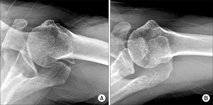
Fig. 1 A meso-acromion (A) and pre-acromion (B) on axial radiograph.
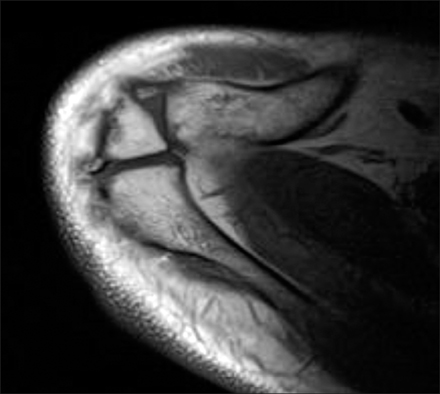
Fig. 2 The fusion defect was seen on the axial T1 magnetic resonance image.
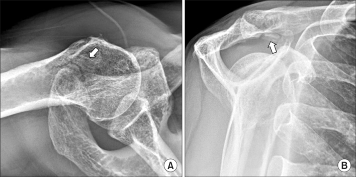
Fig. 3 (A) Axial radiograph giving a false impression of pre-acromion (arrow) (B) Y view of the same patient, clearly showing a fractured acromion spur (arrow).
Reference
-
1. Edelson JG, Zuckerman J, Hershkovitz I. Os acromiale: anatomy and surgical implications. J Bone Joint Surg Br. 1993; 75(4):551–555.
Article2. Macalister A. Notes on acromion. J Anat Physiol. 1893; 27(Pt 2):244.1–251.3. Sammarco VJ. Os acromiale: frequency, anatomy, and clinical implications. J Bone Joint Surg Am. 2000; 82(3):394–400.
Article4. Hutchinson MR, Veenstra MA. Arthroscopic decompression of shoulder impingement secondary to Os acromiale. Arthroscopy. 1993; 9(1):28–32.
Article5. Warner JJ, Beim GM, Higgins L. The treatment of symptomatic os acromiale. J Bone Joint Surg Am. 1998; 80(9):1320–1326.
Article6. Wright RW, Heller MA, Quick DC, Buss DD. Arthroscopic decompression for impingement syndrome secondary to an unstable os acromiale. Arthroscopy. 2000; 16(6):595–599.
Article7. Richards AM, Curtis MJ. Fracture of an os acromiale with associated rupture of the coracoclavicular ligaments. J Shoulder Elbow Surg. 2008; 17(6):e8–e11.
Article8. Andrews JR, Byrd JW, Kupferman SP, Angelo RL. The profile view of the acromion. Clin Orthop Relat Res. 1991; (263):142–146.
Article9. Park JG, Lee JK, Phelps CT. Os acromiale associated with rotator cuff impingement: MR imaging of the shoulder. Radiology. 1994; 193(1):255–257.
Article10. Uri DS, Kneeland JB, Herzog R. Os acromiale: evaluation of markers for identification on sagittal and coronal oblique MR images. Skeletal Radiol. 1997; 26(1):31–34.
Article11. Lee DH, Lee KH, Lopez-Ben R, Bradley EL. The double-density sign: a radiographic finding suggestive of an os acromiale. J Bone Joint Surg Am. 2004; 86(12):2666–2670.12. Nicholson GP, Goodman DA, Flatow EL, Bigliani LU. The acromion: morphologic condition and age-related changes. A study of 420 scapulas. J Shoulder Elbow Surg. 1996; 5(1):1–11.
Article13. Case DT, Burnett SE, Nielsen T. Os acromiale: population differences and their etiological significance. Homo. 2006; 57(1):1–18.
Article14. Kurtz CA, Humble BJ, Rodosky MW, Sekiya JK. Symptomatic os acromiale. J Am Acad Orthop Surg. 2006; 14(1):12–19.
Article15. Grasso A. The incidence and role of the os acromiale in the acromiohumeral impingement syndrome. Radiol Med. 1992; 84(5):567–570.16. Liberson F. Os acromiale: a contested anomaly. J Bone Joint Surg Am. 1937; 19(3):683–689.17. Coskun N, Karaali K, Cevikol C, Demirel BM, Sindel M. Anatomical basics and variations of the scapula in Turkish adults. Saudi Med J. 2006; 27(9):1320–1325.18. Boehm TD, Rolf O, Martetschlaeger F, Kenn W, Gohlke F. Rotator cuff tears associated with os acromiale. Acta Orthop. 2005; 76(2):241–244.
Article19. Ouellette H, Thomas BJ, Kassarjian A, et al. Re-examining the association of os acromiale with supraspinatus and infraspinatus tears. Skeletal Radiol. 2007; 36(9):835–839.
Article20. Burbank KM, Lemos MJ, Bell G, Lemos DW. Incidence of os acromiale in patients with shoulder pain. Am J Orthop (Belle Mead NJ). 2007; 36(3):153–155.21. Neer CS 2nd. Impingement lesions. Clin Orthop Relat Res. 1983; (173):70–77.
Article
- Full Text Links
-
- Actions
-
Cited
- CITED
-
- Close
- Share
-
- Similar articles
-
- Reverse shoulder arthroplasty with os acromiale
- Operative Treatment of Symptomatic Os Acromiale
- Shoulder Impingement Syndrome: Evaluation of the Causes with MRI
- Whatever Your Preference Is for the Treatment of the Proximal Humeral Fracture
- A Survey on Clinical Characteristics of Patients Visiting Pain Clinics
