Evaluation of Associated Carpal Bone Fractures in Distal Radial Fractures
- Affiliations
-
- 1Department of Orthopedic Surgery, Konyang University College of Medicine, Daejeon, Korea. Valeeno1@kyuh.co.kr
- KMID: 1705529
- DOI: http://doi.org/10.4055/cios.2013.5.2.98
Abstract
- BACKGROUND
The purpose of this study was to investigate the frequency and distribution of associated carpal bone fractures (CBFs) in distal radial fractures (DRFs).
METHODS
Three hundred and thirteen patients who underwent surgical treatment for DRFs between March 2007 and January 2010 were reviewed retrospectively. In this study, 223 patients who had preoperative computed tomography (CT) were included. We investigated the frequency and distribution of associated CBFs on CT scans. The relationship between the frequency of associated CBFs and patient factors such as age, gender, body mass index, and the mechanism of injury was assessed.
RESULTS
CBFs were complicated in 46 of 223 DRFs (20.9%). The distribution of CBFs was 23 cases in the triquetrum, 16 in the lunate, 12 in the scaphoid, five in the hamate, and four in the pisiform. Among the 46 cases, a fracture of one carpal bone occurred in 36 cases, two in seven cases, three in two cases, and four in one case. In 10 of the 46 cases, associated CBFs occurred in more than two carpal bones. No significant differences were observed for age, sex, body mass index, or the mechanism of injury between patients with DRFs and CBFs and those without CBFs.
CONCLUSIONS
Because CBFs that mainly occur in the proximal carpal row are complicated in DRFs at a relatively high frequency, assessment of carpal bones using CT scans is beneficial.
Keyword
MeSH Terms
Figure
-
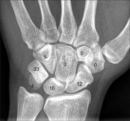
Fig. 1 The distribution of carpal bone fractures that occurred simultaneously with distal radial fractures.

Fig. 2 Initial radiographs (A and B) show a distal radial fracture (AO type C2) and an ulnar styloid fracture. Reconstructed computed tomography images (C and D) show a palmar pole fracture of the lunate. AO: arbeitsgemeinschaft für osteosyntheses.

Fig. 3 Initial radiographs (A and B) show a radial fracture (AO type C3) and an ulnar styloid fracture. A small bony fragment was identified on a lateral radiograph. Reconstructed axial computed tomography image (C) shows a dorsal cortical fracture of the triquetrum and a sagittal fracture of the pisiform. AO: arbeitsgemeinschaft für osteosyntheses.
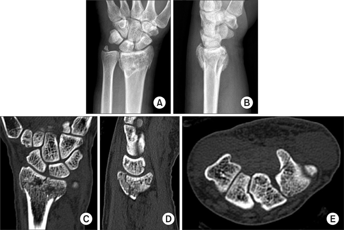
Fig. 4 Initial radiographs (A and B) show a distal radial fracture (AO type C3) and an ulnar styloid fracture. However, no carpal bone fracture is shown on simple radiographs. Reconstructed computed tomography images show fractures of the scaphoid waist (C), palmar pole of the lunate (D), and the hook of the hamate (E). AO: arbeitsgemeinschaft für osteosyntheses.

Fig. 5 Initial radiographs (A and B) show a distal radial fracture (AO type C3) and an ulnar styloid fracture. Bony fragments of the distal radial fracture are displaced over the dorsum of the lunate, and a small bony fragment was identified on a lateral radiograph. Reconstructed computed tomography images show a dorsal pole fracture of the scaphoid (C), a cartilage injury of the dorsal pole of the lunate (D), a dorsal cortical fracture of the triquetrum, and a comminuted fracture of the pisiform (E). AO: arbeitsgemeinschaft für osteosyntheses.
Cited by 2 articles
-
Concomitant Carpal Injuries in Distal Radius Fractures: Retrospective Analysis by Plain Radiographs and Computed Tomography
Chul-Hyun Cho, Eun-Seok Son
J Korean Fract Soc. 2015;28(1):1-7. doi: 10.12671/jkfs.2015.28.1.1.Comparison of Distal Radius Fractures with or without Scaphoid Fractures
Jin Rok Oh, Dong Woo Lee, Jun Pyo Lee
J Korean Soc Surg Hand. 2016;21(1):23-28. doi: 10.12790/jkssh.2016.21.1.23.
Reference
-
1. Osterman AL, VanDuzer ST. Arthroscopy in the treatment of distal radial fractures with assessment and treatment of associated injuries. Atlas Hand Clin. 2006. 11(2):231–241.2. Lindau T, Arner M, Hagberg L. Intraarticular lesions in distal fractures of the radius in young adults: a descriptive arthroscopic study in 50 patients. J Hand Surg Br. 1997. 22(5):638–643.3. Geissler WB, Freeland AE, Savoie FH, McIntyre LW, Whipple TL. Intracarpal soft-tissue lesions associated with an intra-articular fracture of the distal end of the radius. J Bone Joint Surg Am. 1996. 78(3):357–365.4. Turner RG, Faber KJ, Athwal GS. Complications of distal radius fractures. Hand Clin. 2010. 26(1):85–96.5. Wolfe SW. Wolfe SW, Hotchkiss RN, Pederson WC, Kozin SH, editors. Distal radius fractures. Green's operative hand surgery. 2011. 6th ed. Philadelphia, PA: Churchill Livingstone;561–638.6. Pretell-Mazzini J, Carrigan RB. Simultaneous distal radial fractures and carpal bones injuries in children: a review article. J Pediatr Orthop B. 2011. 20(5):330–333.7. Muller ME, Nazarian S, Koch P, Schatzker J. The comprehensive classification of fractures long bones. 1990. New York: Springer-Verlag;54–63.8. Vigler M, Aviles A, Lee SK. Carpal fractures excluding the scaphoid. Hand Clin. 2006. 22(4):501–516.9. Papp S. Carpal bone fractures. Hand Clin. 2010. 26(1):119–127.10. Welling RD, Jacobson JA, Jamadar DA, Chong S, Caoili EM, Jebson PJ. MDCT and radiography of wrist fractures: radiographic sensitivity and fracture patterns. AJR Am J Roentgenol. 2008. 190(1):10–16.11. Kiuru MJ, Haapamaki VV, Koivikko MP, Koskinen SK. Wrist injuries; diagnosis with multidetector CT. Emerg Radiol. 2004. 10(4):182–185.12. van Onselen EB, Karim RB, Hage JJ, Ritt MJ. Prevalence and distribution of hand fractures. J Hand Surg Br. 2003. 28(5):491–495.13. Hove LM. Fractures of the hand: distribution and relative incidence. Scand J Plast Reconstr Surg Hand Surg. 1993. 27(4):317–319.14. Trumble TE, Benirschke SK, Vedder NB. Ipsilateral fractures of the scaphoid and radius. J Hand Surg Am. 1993. 18(1):8–14.15. Rutgers M, Mudgal CS, Shin R. Combined fractures of the distal radius and scaphoid. J Hand Surg Eur Vol. 2008. 33(4):478–483.16. Carter PR, Frederick HA, Laseter GF. Open reduction and internal fixation of unstable distal radius fractures with a low-profile plate: a multicenter study of 73 fractures. J Hand Surg Am. 1998. 23(2):300–307.17. Smith DW, Brou KE, Henry MH. Early active rehabilitation for operatively stabilized distal radius fractures. J Hand Ther. 2004. 17(1):43–49.18. Lozano-Calderon SA, Souer S, Mudgal C, Jupiter JB, Ring D. Wrist mobilization following volar plate fixation of fractures of the distal part of the radius. J Bone Joint Surg Am. 2008. 90(6):1297–1304.19. Smith DK, Murray PM. Avulsion fractures of the volar aspect of triquetral bone of the wrist: a subtle sign of carpal ligament injury. AJR Am J Roentgenol. 1996. 166(3):609–614.20. Cockshott WP. Distal avulsion fractures of the scaphoid. Br J Radiol. 1980. 53(635):1037–1040.21. Slutsky DJ. Predicting the outcome of distal radius fractures. Hand Clin. 2005. 21(3):289–294.
- Full Text Links
-
- Actions
-
Cited
- CITED
-
- Close
- Share
-
- Similar articles
-
- Carpal Bone Fractures in Distal Radial Fractures: Is Computed Tomography Expedient?
- Concomitant Carpal Injuries in Distal Radius Fractures: Retrospective Analysis by Plain Radiographs and Computed Tomography
- Radiologic Follow-up Results of Distraction After Treatment of Distal Radius Fractures using External Fixator
- Operative Treatment of Carpal Scaphoid Fractures with Herbert Screw
- Pediatric Fractures around the Wrist
