Retropharyngeal Spindle Cell/Pleomorphic Lipoma
- Affiliations
-
- 1Department of Radiology, Chonbuk National University Medical School and Hospital, Jeonju 561-712, Korea. sbh1010@jbnu.ac.kr
- 2Department of Otolaryngology-Head and Neck Surgery, Chonbuk National University Medical School and Hospital, Jeonju 561-712, Korea.
- 3Department of Pathology, Chonbuk National University Medical School and Hospital, Jeonju 561-712, Korea.
- KMID: 1705465
- DOI: http://doi.org/10.3348/kjr.2013.14.3.493
Abstract
- Spindle cell/pleomorphic lipoma is an uncommon benign adipose tissue tumor most frequently arising from the subcutaneous tissue of the back, shoulder, head and neck, and extremities. The deep cervical spaces are the rarely affected locations. Herein we report on the imaging findings of spindle cell/pleomorphic lipoma involving the retropharyngeal space in an elderly woman.
Keyword
MeSH Terms
Figure
-
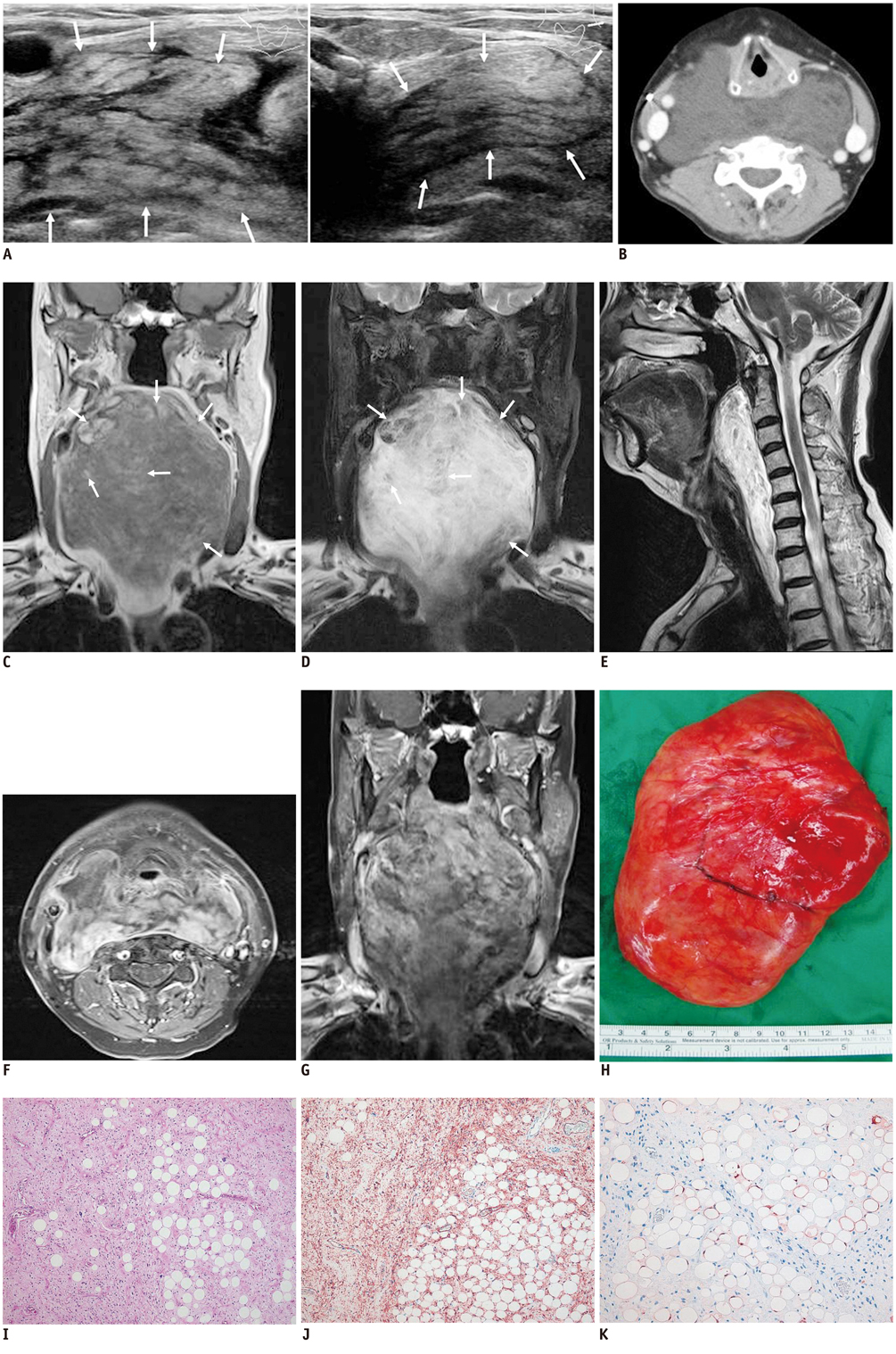
Fig. 1 Retropharyngeal spindle cell/pleomorphic lipoma in 69-year-old woman. A. Ultrasonography (left: right side of neck, right: left side of neck) shows large heterogeneous mass (arrows) in both lateral sides of neck, and diffuse hyperechogenecity with multiple linear or band-shaped hypoechoic portions. B. Contrast-enhanced axial CT image shows huge and relatively well defined, minimal enhancing mass, including multifocal low attenuated areas (-15 to -40 Hounsfield unit). Mass is located in retropharyngeal space extending to both lateral sides of neck. Adjacent structures are displaced by tumor, with no evidence of invasion. C-G. On magnetic resonance imaging, mass is observed to have diffusely hypointense signal on T1-weighted image (WI) (C) and hyperintense signal on fat saturated T2WI (D). There are multifocal areas of high signal intensity within mass on T1WI which showed signal loss on fat saturated T2WI (arrows). Mass extends up to soft palate level cranially and thoracic inlet level caudally on sagittal T2WI (E). Fat saturated gadolinium-enhanced axial (F) and coronal (G) T1WI show exceedingly heterogeneous enhancing mass. H. Surgical specimen shows well-circumscribed and lobulated mass with thin fibrous capsule. I. Most of tumor contains spindle shaped cells in collagenous background. Between spindle cells, mature adipose tissue and characteristic floret-like multinucleated cells are present (Hematoxylin & Eosin staining; original magnification, × 10). J. Spindle tumor cells are positive for CD34 (immunohistochemical staining; original magnification, × 10). K. Mature fat cells are positive for S-100 protein. In contrast, spindle cells have no immunoreactivity for S-100 protein (immunohistochemical staining; original magnification, × 10).
Reference
-
1. French CA, Mentzel T, Kutzner H, Fletcher CD. Intradermal spindle cell/pleomorphic lipoma: a distinct subset. Am J Dermatopathol. 2000. 22:496–502.2. Reis-Filho JS, Milanezi F, Soares MF, Fillus-Neto J, Schmitt FC. Intradermal spindle cell/pleomorphic lipoma of the vulva: case report and review of the literature. J Cutan Pathol. 2002. 29:59–62.3. Vecchio G, Amico P, Caltabiano R, Colella G, Lanzafame S, Magro G. Spindle cell/pleomorphic lipoma of the oral cavity. J Craniofac Surg. 2009. 20:1992–1994.4. Gu MJ, Sohn KR, Park JH. Spindle cell/pleomorphic lipoma of the oropharynx. Korean J Pathol. 2009. 43:580–582.5. Gurel D, Kargi A, Lebe B. Pedunculated cutaneous spindle cell/pleomorphic lipoma. J Cutan Pathol. 2010. 37:e57–e59.6. Dal Cin P, Sciot R, Polito P, Stas M, de Wever I, Cornelis A, et al. Lesions of 13q may occur independently of deletion of 16q in spindle cell/pleomorphic lipomas. Histopathology. 1997. 31:222–225.7. Mandahl N, Mertens F, Willén H, Rydholm A, Brosjö O, Mitelman F. A new cytogenetic subgroup in lipomas: loss of chromosome 16 material in spindle cell and pleomorphic lipomas. J Cancer Res Clin Oncol. 1994. 120:707–711.8. Bancroft LW, Kransdorf MJ, Peterson JJ, Sundaram M, Murphey MD, O'Connor MI. Imaging characteristics of spindle cell lipoma. AJR Am J Roentgenol. 2003. 181:1251–1254.9. Chong VF, Fan YF. Radiology of the retropharyngeal space. Clin Radiol. 2000. 55:740–748.
