Caruncular Abscess Due to Actinomycosis
- Affiliations
-
- 1Department of Ophthalmology, Chungnam National University College of Medicine, Daejeon, Korea. sblee@cnu.ac.kr
- KMID: 1705426
- DOI: http://doi.org/10.3341/kjo.2013.27.4.288
Abstract
- The authors report a caruncular abscess caused by actinomycosis. A 47-year-old woman was admitted with persistent purulent discharge from the caruncle of the left eye for a duration of six months. Excisional drainage was performed, and 'sulfur granules' were observed, consistent with actinomyces infection. Intraoperative lacrimal probing and irrigation were performed to confirm that the abscess and canaliculus were not connected. Oral and topical antibiotics were administered postoperatively; the lesion resolved with no evidence of recurrence, and the symptom improved.
Keyword
MeSH Terms
Figure
-
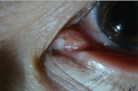
Fig. 1 The slit lamp photograph shows swelling and erythema in the left pericaruncular area, with purulent drainage.
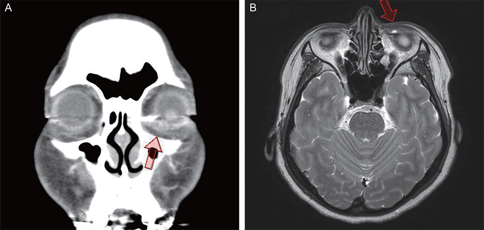
Fig. 2 (A) Orbital computed tomography. Peripheral enhancing low-density lesion, approximately 1 cm in diameter, in the left lower eyelid area, suggestive of an abscess. (B) T2-weighted magnetic resonance imaging shows a high signal intensity lesion of the same size in the medial portion of the left lower eyelid area, consistent with an abscess.
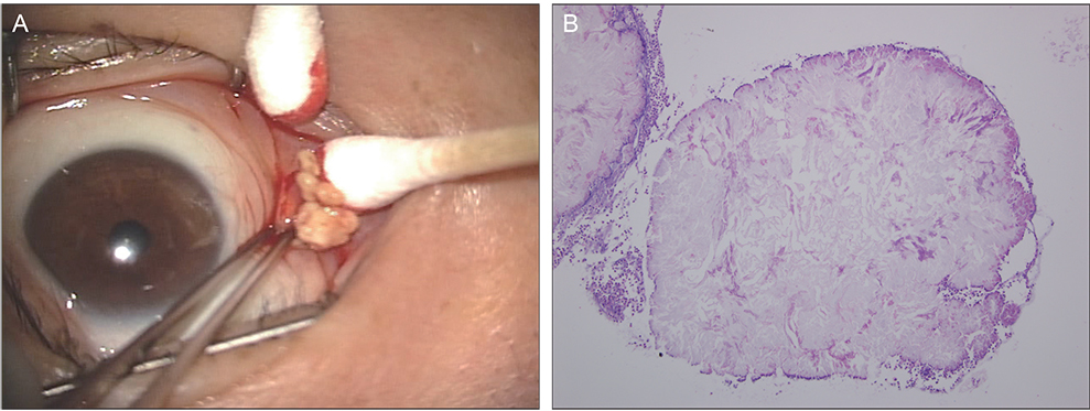
Fig. 3 (A) Intraoperative microscopic photograph shows 'sulfur granules' in the caruncular cyst. (B) Histopathologically, actinomycotic sulfur granules are identified. The bulk of the granule is composed of amorphous material with neutrophils clinging to the edge (hematoxylin-eosin, ×100).
Reference
-
1. Luthra CL, Doxanas MT, Green WR. Lesions of the caruncle: a clinicohistopathologic study. Surv Ophthalmol. 1978; 23:183–195.2. Shields CL, Shields JA, White D, Augsburger JJ. Types and frequency of lesions of the caruncle. Am J Ophthalmol. 1986; 102:771–778.3. Santos A, Gomez-Leal A. Lesions of the lacrimal caruncle. Clinicopathologic features. Ophthalmology. 1994; 101:943–949.4. Koo L, Chang EL. Methicillin-resistant Staphylococcus aureus caruncle abscess. Ophthal Plast Reconstr Surg. 2007; 23:160–161.5. Pappalardo J, Lee GA, Whitehead K. Actinomycotic granule of the caruncle: a case report. Ophthal Plast Reconstr Surg. 2011; 27:e100–e102.6. Briscoe D, Edelstein E, Zacharopoulos I, et al. Actinomyces canaliculitis: diagnosis of a masquerading disease. Graefes Arch Clin Exp Ophthalmol. 2004; 242:682–686.7. Sullivan TJ, Aylward GW, Wright JE. Actinomycosis of the orbit. Br J Ophthalmol. 1992; 76:505–506.8. Roussel TJ, Olson ER, Rice T, et al. Chronic postoperative endophthalmitis associated with Actinomyces species. Arch Ophthalmol. 1991; 109:60–62.
- Full Text Links
-
- Actions
-
Cited
- CITED
-
- Close
- Share
-
- Similar articles
-
- Destruction of the C2 Body due to Cervical Actinomycosis: Connection between Spinal Epidural Abscess and Retropharyngeal Abscess
- Submucosal Gastric Actinomycosis in a Hematemesis Patient
- Actinomycosis Presented as Acute Appendicitis
- Vertebral Actinomycosis: Two Case Report
- A Case Of Pelvic Actinomycosis Complicated By Tuboovarian Abscess
