Korean J Pediatr Infect Dis.
2014 Apr;21(1):59-64.
A Case of Subdural Empyema Caused by Sinusitis in a Child
- Affiliations
-
- 1Department of Pediatrics, Yonsei University College of Medicine, Seoul, Korea.
- 2Department of Neurosurgery, Yonsei University College of Medicine, Seoul, Korea.
- 3Department of Otorhinolaryngology, Yonsei University College of Medicine, Seoul, Korea.
- 4Department of Pediatrics, National Health Insurance Service Ilsan Hospital, Goyang, Korea. janggwangc@yuhs.ac
Abstract
- The current paper reports on a case of subdural empyema secondary to frontal sinusitis in an otherwise healthy child. Sinusitis is a common and benign condition in most pediatric cases. Because of the widespread use of antibiotics, intracranial extension of pediatric sinusitis is rarely seen today; however, complications (e.g., cavernous sinus thrombosis, orbital infection, meningitis, and subdural empyema) are potentially life threatening. A 15-year-old right-handed male presented with a 3-day history of fever, headache, and left-sided palsy. Computed tomography revealed right-sided subdural empyema with right frontal sinusitis and maxillary sinusitis. A postoperative inpatient neurological consultation was requested 2 months post-surgery due to motor function deficits. The results suggested that early and accurate diagnosis of subdural empyema leads to prompt treatment and a favorable outcome for the patient.
MeSH Terms
Figure
-
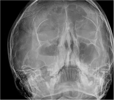
Fig. 1 Waters' view demonstrating right acute maxillary sinusitis & frontal sinusitis with an air-fluid level.
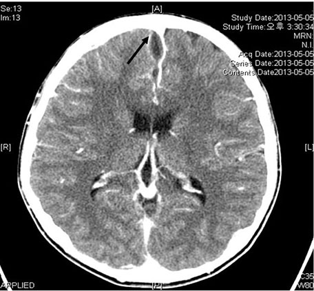
Fig. 2 Axial contrast-enhanced brain CT scan demonstrating small amount of subdural fluid with meningeal enhancement along the anterior falx and right frontal convexity (arrow).
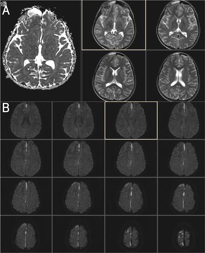
Fig. 3 T1 & T2 weighted contrast cerebral MRI showed a thin layer of subdural collection along falx and interhemispheric fissure with restricted diffusion (typical finding of subdural empyema). (A) T2 weighted contrast cerebral MRI showed meningitis extends over right cerebral hemisphere, probably direct extension from sinusitis. (B) T1 weighted contrast cerebral MRI showed multifocal empyemas in right frontal region and along the falx cerebri.
Reference
-
1. Mackin LA, Antonini CJ Jr. Acute sinusitis. Lippincotts Prim Care Pract. 1999; 3:65–69.2. McQuillan L, Crane LA, Kempe A. Diagnosis and management of acute sinusitis by pediatricians. Pediatrics. 2009; 123:e193–e198.
Article3. Isaacson G. Sinusitis in childhood. Pediatr Clin North Am. 1996; 43:1297–1318.
Article4. DeMuri GP, Wald ER. Complications of acute bacterial sinusitis in children. Pediatr Infect Dis J. 2011; 30:701–702.
Article5. Morice AH, Fontana GA, Sovijarvi AR, Pistolesi M, Chung KF, Widdicombe J, et al. The diagnosis and management of chronic cough. Eur Respir J. 2004; 24:481–492.
Article6. Barbi E, Longo G. Chronic and recurrent cough, sinusitis and asthma. Much ado about nothing. Pediatr Allergy Immunol. 2007; 18:Suppl 18. 22–24.
Article7. Yun DJ, Hong CH, Oh KK. Chronic cough and sinusitis in children--the role of antimicrobials. Yonsei Med J. 1983; 24:67–75.
Article8. Welch JE, Hogan MB, Wilson NW. Ten-year experience using a plastic, disposable curette for the diagnosis of primary ciliary dyskinesia. Ann Allergy Asthma Immunol. 2004; 93:189–192.
Article9. Kombogiorgas D, Seth R, Athwal R, Modha J, Singh J. Suppurative intracranial complications of sinusitis in adolescence. Single institute experience and review of literature. Br J Neurosurg. 2007; 21:603–609.
Article10. DeMuri GP, Wald ER. Clinical practice. Acute bacterial sinusitis in children. N Engl J Med. 2012; 367:1128–1134.11. Holland AA, Morriss M, Glasier PC, Stavinoha PL. Complicated subdural empyema in an adolescent. Arch Clin Neuropsychol. 2013; 28:81–91.
Article12. Chow AW, Benninger MS, Brook I, Brozek JL, Goldstein EJ, Hicks LA, et al. IDSA clinical practice guideline for acute bacterial rhinosinusitis in children and adults. Clin Infect Dis. 2012; 54:e72–e112.
Article13. Betz CS, Issing W, Matschke J, Kremer A, Uhl E, Leunig A. Complications of acute frontal sinusitis: a retrospective study. Eur Arch Otorhinolaryngol. 2008; 265:63–72.
Article14. Gupta S, Vachhrajani S, Kulkarni AV, Taylor MD, Dirks P, Drake JM, et al. Neurosurgical management of extraaxial central nervous system infections in children. J Neurosurg Pediatr. 2011; 7:441–451.
Article
- Full Text Links
-
- Actions
-
Cited
- CITED
-
- Close
- Share
-
- Similar articles
-
- Subdural Empyema Concomitant with Bilateral Subdural Effusion in Infant after Meningitis
- A Case of Subtentorial Subdural Empyema Resulting from Chronic Otitis Media with Cholesteatoma
- Fatal Subdural Empyema Following Pyogenic Meningitis
- Two Cases of Intracranial Subdural Empyema
- A Case of Subdural Empyema Caused by Escherichia coli Infection
