Obstet Gynecol Sci.
2014 May;57(3):181-186. 10.5468/ogs.2014.57.3.181.
Clinical characteristics and outcomes of antenatal fetal intra-abdominal umbilical vein varix detection
- Affiliations
-
- 1Department of Obstetrics and Gynecology, Cheil General Hospital and Women's Healthcare Center, Kwandong University College of Medicine, Seoul, Korea. mykimdr@yahoo.co.kr
- 2Department of Obstetrics and Gynecology, Woosung Woman's Hospital, Ansan, Korea.
- KMID: 1704423
- DOI: http://doi.org/10.5468/ogs.2014.57.3.181
Abstract
OBJECTIVE
This study reviewed clinical characteristics of fetal intra-abdominal umbilical vein (FIUV) varices that were detected during antenatal ultrasound examinations.
METHODS
Between January 2006 and January 2012, 121 cases of FIUV varices were detected and 7 cases were lost to follow-up. We retrospectively reviewed the medical records of 114 patients and neonates.
RESULTS
From a total 96,553 ultrasound examinations in 43,995 pregnancies, 121 cases of FIUV varices were identified (2.8 per 1,000 pregnancies). Gestational age at diagnosis was 32.0 +/- 2.9 weeks (range, 20.1-36.3 weeks), the mean diameter of the FIUV varix was 12.6 +/- 2.1 mm (range, 8.0-21.0 mm) at initial diagnosis and the mean maximal diameter was 13.1 +/- 2.3 mm (range, 8.0-21.0 mm) during follow-up. The most severe pregnancy complications included one case of intrauterine fetal death and another case of fetal hydrops. Associated fetal anomalies (n = 11, 9.6%) detected by ultrasonography included bilateral renal pelvis dilatation, ventriculomegaly, cryptorchidism, incomplete renal duplication and pulmonary sequestration. A total of 104 cases (91.2%) were delivered at term and 10 cases (8.8%) were preterm deliveries before 37 weeks of gestation.
CONCLUSION
FIUV varices that are not associated with fetal anomalies based on ultrasound examination during prenatal care have favorable pregnancy outcomes. Nevertheless, close fetal monitoring is recommended to decrease perinatal complications.
MeSH Terms
-
Bronchopulmonary Sequestration
Cryptorchidism
Diagnosis
Dilatation
Female
Fetal Death
Fetal Monitoring
Follow-Up Studies
Gestational Age
Humans
Hydrops Fetalis
Infant, Newborn
Kidney Pelvis
Lost to Follow-Up
Male
Medical Records
Pregnancy
Pregnancy Complications
Pregnancy Outcome
Prenatal Care
Retrospective Studies
Ultrasonography
Umbilical Veins*
Varicose Veins*
Figure
-
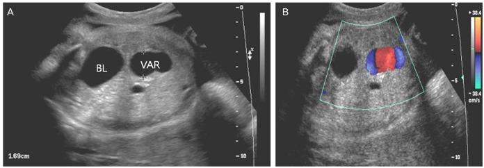
Fig. 1 (A) Transverse view of the lower fetal abdomen showing an umbilical vein varix that was approximately 16.9 mm at 33 weeks of gestation. (B) Color Doppler shows some turbulence in the intravascular area and differentiates from other cystic lesions. BL, bladder; VAR, fetal umbilical vein varix.
Cited by 1 articles
-
A Huge Umbilical Vein Aneurysm: Case Report and a Brief Review of Literatures Describing Umbilical Vessel Aneurysm
Jae Hoon Lee, Ju Hyun Cho, Ha Yan Kwon, Yong Won Park, Young Han Kim
Korean J Perinatol. 2014;25(3):178-183. doi: 10.14734/kjp.2014.25.3.178.
Reference
-
1. Mantas N, Sifakis S, Koukoura O, Avgoustinakis E, Koumantakis E. Intraabdominal umbilical vein dilatation and term delivery: a case report and review of the literature. Fetal Diagn Ther. 2007; 22:431–434. PMID: 17652931.2. Nyberg D. Varix of the umbilical vein. In : Nyberg DA, McGahan JP, Pretorius DH, Pilu G, editors. Diagnostic imaging of fetal anomalies. Philadelphia: Lippincott Williams & Wilkins;2003. p. 114–115.3. Weissman A, Jakobi P, Bronshtein M, Goldstein I. Sonographic measurements of the umbilical cord and vessels during normal pregnancies. J Ultrasound Med. 1994; 13:11–14. PMID: 7636947.
Article4. Mahony BS, McGahan JP, Nyberg DA, Reisner DP. Varix of the fetal intra-abdominal umbilical vein: comparison with normal. J Ultrasound Med. 1992; 11:73–76. PMID: 1560496.
Article5. Sepulveda W, Mackenna A, Sanchez J, Corral E, Carstens E. Fetal prognosis in varix of the intrafetal umbilical vein. J Ultrasound Med. 1998; 17:171–175. PMID: 9514169.
Article6. Rahemtullah A, Lieberman E, Benson C, Norton ME. Outcome of pregnancy after prenatal diagnosis of umbilical vein varix. J Ultrasound Med. 2001; 20:135–139. PMID: 11211133.
Article7. Byers BD, Goharkhay N, Mateus J, Ward KK, Munn MB, Wen TS. Pregnancy outcome after ultrasound diagnosis of fetal intra-abdominal umbilical vein varix. Ultrasound Obstet Gynecol. 2009; 33:282–286. PMID: 19115263.
Article8. Weissmann-Brenner A, Simchen MJ, Moran O, Kassif E, Achiron R, Zalel Y. Isolated fetal umbilical vein varix: prenatal sonographic diagnosis and suggested management. Prenat Diagn. 2009; 29:229–233. PMID: 19177454.9. Fung TY, Leung TN, Leung TY, Lau TK. Fetal intra-abdominal umbilical vein varix: what is the clinical significance? Ultrasound Obstet Gynecol. 2005; 25:149–154. PMID: 15685644.
Article10. Zalel Y, Lehavi O, Heifetz S, Aizenstein O, Dolitzki M, Lipitz S, et al. Varix of the fetal intra-abdominal umbilical vein: prenatal sonographic diagnosis and suggested in utero management. Ultrasound Obstet Gynecol. 2000; 16:476–478. PMID: 11169334.
- Full Text Links
-
- Actions
-
Cited
- CITED
-
- Close
- Share
-
- Similar articles
-
- Fetal Intra-abdominal Umbilical Vein Varix Complicated with Patent Ductus Venosus and Atrial Septal Defect
- A Case of Varix of the Fetal Intra-Abdominal Umbilical Vein and Review of the Literature
- A Case Report: Varix of the Intrafetal Umbilical Vein
- A Huge Umbilical Vein Aneurysm: Case Report and a Brief Review of Literatures Describing Umbilical Vessel Aneurysm
- A case of one fetal demise of twin pregnancy by umbilical artery stricture
