A Case of Monophasic Fibrous Synovial Sarcoma Confirmed Primary Pulmonary Origin by 18F-FDG PET/CT
- Affiliations
-
- 1Division of Pulmonary and Critical Care Medicine, Department of Internal Medicine, Kangbuk Samsung Hospital, Sungkyunkwan University School of Medicine, Seoul, Korea. mdlimsy@skku.edu
- 2Department of Radiology, Kangbuk Samsung Hospital, Sungkyunkwan University School of Medicine, Seoul, Korea.
- 3Department of Pathology, Kangbuk Samsung Hospital, Sungkyunkwan University School of Medicine, Seoul, Korea.
- 4Department of Thoracic Surgery, Kangbuk Samsung Hospital, Sungkyunkwan University School of Medicine, Seoul, Korea.
- KMID: 1630806
- DOI: http://doi.org/10.4046/trd.2006.60.6.673
Abstract
- Most malignant mesenchymal tumors of the lung are metastases of a primary tumor from elsewhere in the body. A primary pulmonary synovial sarcoma is a very rare neoplasm that accounts for approximately 10% of soft tissue sarcomas and makes up only 0.5% of all primary lung malignancies. We report a case of a primary pulmonary synovial sarcoma in a 60-year old woman. In this case, a lung metastasis was excluded using 18F-FDG PET /CT imaging.
MeSH Terms
Figure
-

Figure 1 (A) Chest radiograph shows right lower lobe mass. (B) On chest CT scan, 5×4cm sized well demarcated and highly enhancing mass was noted in right lower lobe.

Figure 2 (A) FDG PET coronal image: FDG PET scan reveals increased FDG uptake foci in right lower lobe (peak SUV=3.7). (B) FDG PET fused coronal image: fusion PET demonstrated mild hypermetabolic mass in right lower lobe.

Figure 3 The cut surface of the resected lung shows a relatively well demarcated pale yellow tan solid mass with fish flesh-like appearance. The bronchial tree is grossly intact.
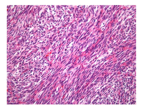
Figure 4 Light microscopic finding shows that tumor is composed of highly cellular spindle cells with long intersecting fascicular arrangement and mild to moderate atypism (H&E, ×400).

Figure 5 The results of immunohistochemical stains. The tumor cells are completely negative for smooth muscle actin (A; ×400) and CD34 (B; ×400). The majority of tumor cells show strong and diffuse positive reaction for bcl-2 (C; ×400) and vimentin (D; ×400).
Reference
-
1. Hosono T, Hironaka M, Kobayashi A, Yamasawa H, Bando M, Ohno S, et al. Primary pulmonary synovial sarcoma confirmed by molecular detection of SYT-SSX1 fusion gene transcripts: a case report and review of the literature. Jpn J Clin Oncol. 2005. 35:274–279.2. Hisaoka M, Hashimoto H, Iwamasa T, Ishikawa K, Aoki T. Primary synovial sarcoma of the lung: report of two cases confirmed by molecular detection of SYT-SSX fusion gene transcripts. Histopathology. 1999. 34:205–210.3. Niwa H, Masuda S, Kobayashi C, Oda Y. Pulmonary synovial sarcoma with polypoid endobronchial growth: a case report, immunohistochemical and cytogenetic study. Pathol Int. 2004. 54:611–615.4. Okamoto S, Hisaoka M, Daa T, Hatakeyama K, Iwamasa T, Hashimoto H. Primary pulmonary synovial sarcoma: a clinicopathologic, immunohistochemical, and molecular study of 11 cases. Hum Pathol. 2004. 35:850–856.5. Chan JA, McMenamin ME, Fletcher CD. Synovial sarcoma in older patients: clinicopathological analysis of 32 cases with emphasis on unusual histological features. Histopathology. 2003. 43:72–83.6. Zeren H, Moran CA, Suster S, Fishback NF, Koss MN. Primary pulmonary sarcomas with features of monophasic synovial sarcoma: a clinicopathological, immunohisto-chemical, and ultrastructural study of 25 cases. Hum Pathol. 1995. 26:474–480.7. Duran-Mendicuti A, Costello P, Vargas SO. Primary synovial sarcoma of the chest: radiographic and clinicopathologic correlation. J Thorac Imaging. 2003. 18:87–93.8. Yoon GS, Park SY, Kang GH, Kim OJ. Primary pulmonary sarcoma with morphologic features of biphasic synovial sarcoma: a case report. J Korean Med Sci. 1998. 13:71–76.9. Song SH, Lee KH, Oh JH, Moon HS, Song JS, Park SH, et al. A case of primary pulmonary sarcoma with morphologic features of biphasic synovial sarcoma. Tuberc Respir Dis. 1998. 45:1284–1289.10. Dennison S, Weppler E, Giacoppe G. Primary pulmonary synovial sarcoma: a case report and review of current diagnostic and therapeutic standards. Oncologist. 2004. 9:339–342.11. Kawai A, Woodruff J, Healey JH, Brennan MF, Antonescu CR, Ladanyi M. SYT-SSX gene fusion as a determinant of morphology and prognosis in synovial sarcoma. N Engl J Med. 1998. 338:153–160.12. Spillane AJ, A'Hern R, Judson IR, Fisher C, Thomas JM. Synovial sarcoma: a clinicopathologic, staging, and prognostic assessment. J Clin Oncol. 2000. 18:3794–3803.
- Full Text Links
-
- Actions
-
Cited
- CITED
-
- Close
- Share
-
- Similar articles
-
- A Case of Primary Pulmonary Artery Sarcoma Mimicking Pulmonary Embolism: Role of PET/CT for Differential Diagnosis
- Use of 18F-FDG PET/CT in Second Primary Cancer
- A Study of 3 Cases of Synovial Sarcoma by Immunohistochemical Stain and Electron Microscopy
- Lung Adenocarcinoma Staged as an Unknown Primary Presenting with Symptomatic Colon Metastases: Staging by 18F-FDG PET/CT
- Recurrent Follicular Dendritic Cell Sarcoma of the Parotid Gland Imaged with 18F-FDG PET/CT
