Korean J Ophthalmol.
2013 Feb;27(1):55-57. 10.3341/kjo.2013.27.1.55.
Conjunctival Hypertrophic Scar Following Cryotherapy for Retinopathy of Prematurity
- Affiliations
-
- 1Department of Ophthalmology, Samsung Medical Center, Sungkyunkwan University School of Medicine, Seoul, Korea. ydkimoph@skku.edu
- 2Department of Ophthalmology, Asan Medical Center, University of Ulsan College of Medicine, Seoul, Korea.
- KMID: 1501798
- DOI: http://doi.org/10.3341/kjo.2013.27.1.55
Abstract
- A 6-year-old boy was referred to our hospital with symblepharon and lateral canthal deformity in both eyes, which developed 6 years ago. The patient was born at 27 weeks gestation. He had received cryotherapy for retinopathy of prematurity. One month after cryotherapy, he developed a conjunctival scar with symblepharon in both eyes and underwent symblepharon lysis at another hospital 5 years prior. Ocular examination revealed an extensive conjunctival hypertrophic scar with symblepharon and limitation of extraocular movements. An excisional biopsy, lateral canthoplasty, and symblepharon lysis with conjunctival autograft from the contralateral eye were performed in the left eye. Histopathologic examination revealed diffuse proliferation and infiltration of collagenous tissue.
MeSH Terms
Figure
-
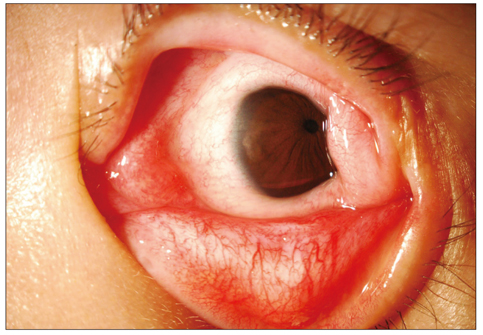
Fig. 1 Anterior segment photographs showing lateral canthal deformity, symblepharon, and a conjunctival scar involving almost 360 degrees of peripheral cornea in the left eye.
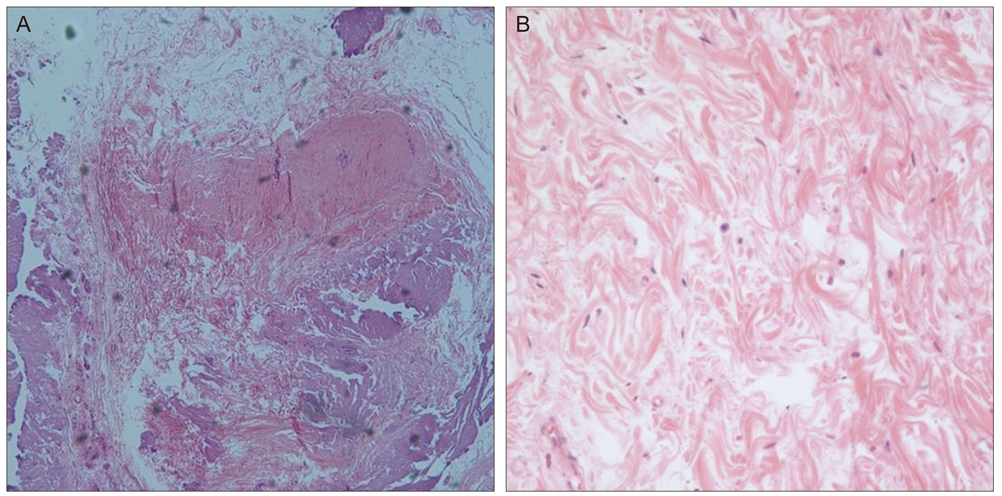
Fig. 2 Histopathologic examination reveals diffuse proliferation and infiltration of the collagenous tissue. These findings are consistent with hypertrophic scar. H&E, (A) ×100, (B) ×400.

Fig. 3 (A) Preoperative photograph showing a temporal conjunctival scar with lateral canthal deformity of both eyes. (B,C) Photograph taken 1 year after surgery showing no recurrence and good cosmesis.
Reference
-
1. Cryotherapy for Retinopathy of Prematurity Cooperative Group. Multicenter trial of cryotherapy for retinopathy of prematurity: preliminary results. Arch Ophthalmol. 1988. 106:471–479.2. Cryotherapy for Retinopathy of Prematurity Cooperative Group. Multicenter trial of cryotherapy for retinopathy of prematurity: Snellen visual acuity and structural outcome at 5 1/2 years after randomization. Arch Ophthalmol. 1996. 114:417–424.3. Cryotherapy for Retinopathy of Prematurity Cooperative Group. Effect of retinal ablative therapy for threshold retinopathy of prematurity: results of Goldmann perimetry at the age of 10 years. Arch Ophthalmol. 2001. 119:1120–1125.4. Brown GC, Tasman WS, Naidoff M, et al. Systemic complications associated with retinal cryoablation for retinopathy of prematurity. Ophthalmology. 1990. 97:855–858.5. Watanabe H, Tsukamoto Y, Saito Y, et al. Massive proliferation of conjunctival tissue after cryotherapy for retinopathy of prematurity. Arch Ophthalmol. 1997. 115:278–279.6. Park JW, Cho HJ, Cho YW. Transscleral diode laser photocoagulation for retinopathy of prematurity: five years' experience. J Korean Ophthalmol Soc. 2006. 47:1960–1965.
- Full Text Links
-
- Actions
-
Cited
- CITED
-
- Close
- Share
-
- Similar articles
-
- Effect of Cryotherapy for Retinopathy of Prematurity
- A Comparison of Cryotherapy Versus Transscleral Diode Laser Photocoagulation for the Treatment of Retinopathy of Prematurity
- Cryotherapy and Myopia in Patients with Retinopathy of Prematurity
- The Effect of Cryotherapy and Laser Photocoagulation for the Retinopathy of Prematurity
- The Effect of Cryotherapy on Stage 3 Retinopathy of Prematurity
