Anatomical Analysis of Clavicles in Korean Adults and Compatibility of Pre-Contoured Anatomical Plates
- Affiliations
-
- 1Department of Orthopedic Surgery, Konkuk University Chungju Hospital, Konkuk University School of Medicine, Chungju, Korea. kodukan@naver.com
- KMID: 1494147
- DOI: http://doi.org/10.4055/jkoa.2013.48.5.350
Abstract
- PURPOSE
Due to the complex anatomy of clavicles, clavicular plates are not always compatible with clavicular fractures. The purpose of this study was to analyze basic data on the anatomy of the clavicle in order to determine compatibility between clavicles of Korean adults and pre-contoured anatomical plates.
MATERIALS AND METHODS
We analyzed the anatomy of 600 clavicles of 300 patients who underwent three-dimensional (3D) computed tomography of clavicles in the emergency room of Konkuk University Chungju Hospital, between July 2010 and July 2011, using Andermahr's method; in addition, the compatibility between 3D axial images of clavicles and sectional images of pre-contoured anatomical plates was also examined using Adobe Photoshop.
RESULTS
The mean length of the clavicle was 146.21+/-4.98 mm, the mean width was 9.63+/-1.67 mm, and the mean thickness was 9.54+/-1.67 mm. The location of the maximum superior bow was 36.17+/-0.60 mm from the lateral end of the clavicle and the mean magnitude was 5.88+/-0.62 mm. The mean depth of medial curvature was 15.89+/-1.33 mm, and the mean depth of the lateral curvature of the clavicle was 11.73+/-1.66 mm. The compatibility between clavicles and plates was 79% as above a fair compatibility in the 50% range of clavicles and 48% as above a fair compatibility in the 60% to 70% range of clavicles. On the contrary, in application of medial and lateral plates in the 60% to 70% range of clavicles, above a fair compatibility had increased to 67%.
CONCLUSION
A more adequate pre-contoured anatomical plate is required for satisfactory improvement of the compatibility of clavicles of Korean adults.
Keyword
MeSH Terms
Figure
-

Figure 1 Measurement of dimensions and curvature of the right clavicle. A, medial edge; B, lateral edge; C, D, anterior edge; E, F, posterior edge; L1, length of the clavicle; L2, depth of the medial curve of the clavicle; L3, depth of the lateral curve of the clavicle; Ant., anterior; Post., posterior; Med., medial; Lat., lateral.

Figure 2 Clavicular width (W; mm) measured at 10% intervals of total length using Aquilion 64 3-dimensional software.

Figure 3 Clavicular thickness (T; mm) measured at 10% interval of total length with using Aquilion 64 3-dimensional software.

Figure 4 Measurement method used to determine the location (D1) and magnitude (D2) of the maximum superior clavicular bow. Measurements were performed using Aquilion 64 3-dimensional software. A, medial edge; B, lateral edge; D1, location of the maximum superior clavicular bow; D2, magnitude of the maximum superior clavicular bow.

Figure 5 Clavicular width (W; mm) measured at 10% interval of total length.
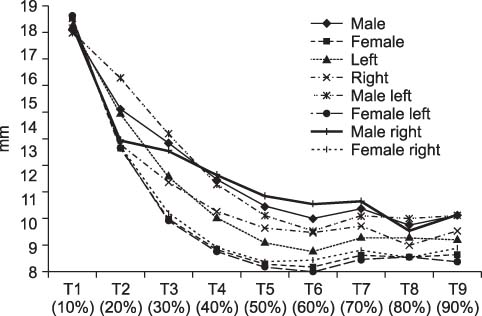
Figure 6 Clavicular thickness (T; mm) measured at 10% interval of total length.

Figure 7 (A) Intra-operative photograph of a clavicle with a plate applied to the medial (Med.) and lateral (Lat.) sides in opposition. (B) Digital images of a clavicle with a plate applied as original. (C) Digital image of a clavicle with a plate applied in a Lat. mid-shaft clavicle fracture. (D) Digital image of a clavicle with a plate applied to the Med. and Lat. sides in opposition. Ant., anterior; Post., posterior.
Reference
-
1. Nordqvist A, Petersson C. The incidence of fractures of the clavicle. Clin Orthop Relat Res. 1994; 300:127–132.
Article2. Moseley HF. The clavicle: its anatomy and function. Clin Orthop Relat Res. 1968; 58:17–27.3. Eskola A, Vainionpää S, Myllynen P, Pätiälä H, Rokkanen P. Surgery for ununited clavicular fracture. Acta Orthop Scand. 1986; 57:366–367.
Article4. Kwon KW, Ahn DJ. A clinical study on surgical treatment of clavicular non-unions. J Korean Orthop Assoc. 1987; 22:1127–1131.
Article5. Neer CS 2nd. Fractures of the distal third of the clavicle. Clin Orthop Relat Res. 1968; 58:43–50.6. Rowe CR. An atlas of anatomy and treatment of midclavicular fractures. Clin Orthop Relat Res. 1968; 58:29–42.7. Wilkins RS, Jjhnston RM. Ununited fractures of the clavicle. J Bone Joint Surg Am. 1983; 65:773–778.
Article8. Hill JM, McGuire MH, Crosby LA. Closed treatment of displaced middle-third fractures of the clavicle gives poor results. J Bone Joint Surg Br. 1997; 79:537–539.
Article9. Zlowodzki M, Zelle BA, Cole PA, Jeray K, McKee MD. Evidence-Based Orthopaedic Trauma Working Group. Treatment of acute midshaft clavicle fractures: systematic review of 2144 fractures: on behalf of the Evidence-Based Orthopaedic Trauma Working Group. J Orthop Trauma. 2005; 19:504–507.10. Iannotti MR, Crosby LA, Stafford P, Grayson G, Goulet R. Effects of plate location and selection on the stability of midshaft clavicle osteotomies: a biomechanical study. J Shoulder Elbow Surg. 2002; 11:457–462.
Article11. Andermahr J, Jubel A, Elsner A, et al. Anatomy of the clavicle and the intramedullary nailing of midclavicular fractures. Clin Anat. 2007; 20:48–56.
Article12. Huang JI, Toogood P, Chen MR, Wilber JH, Cooperman DR. Clavicular anatomy and the applicability of precontoured plates. J Bone Joint Surg Am. 2007; 89:2260–2265.
Article13. Kloen P, Sorkin AT, Rubel IF, Helfet DL. Anteroinferior plating of midshaft clavicular nonunions. J Orthop Trauma. 2002; 16:425–430.
Article14. Mullaji AB, Jupiter JB. Low-contact dynamic compression plating of the clavicle. Injury. 1994; 25:41–45.
Article15. Schwarz N, Höcker K. Osteosynthesis of irreducible fractures of the clavicle with 2.7-MM ASIF plates. J Trauma. 1992; 33:179–183.
Article16. Ferran NA, Hodgson P, Vannet N, Williams R, Evans RO. Locked intramedullary fixation vs plating for displaced and shortened mid-shaft clavicle fractures: a randomized clinical trial. J Shoulder Elbow Surg. 2010; 19:783–789.
Article17. Chandrasenan J, Espag M, Dias R, Clark DI. The use of anatomic precontoured plates in the treatment of midshaft clavicle fractures. J Bone Joint Surg Br. 2009; 91:179–180.
Article18. VanBeek C, Boselli KJ, Cadet ER, Ahmad CS, Levine WN. Precontoured plating of clavicle fractures: decreased hardware-related complications? Clin Orthop Relat Res. 2011; 469:3337–3343.
Article19. Daruwalla ZJ, Courtis P, Fitzpatrick C, Fitzpatrick D, Mullett H. Anatomic variation of the clavicle: A novel three-dimensional study. Clin Anat. 2010; 23:199–209.
Article20. Duprey S, Bruyere K, Verriest JP. Influence of geometrical personalization on the simulation of clavicle fractures. J Biomech. 2008; 41:200–207.
Article
- Full Text Links
-
- Actions
-
Cited
- CITED
-
- Close
- Share
-
- Similar articles
-
- The use of precontoured plates for midshaft clavicle fractures is not always the best course of treatment
- Treatment for Comminuted fractures of Distal End of Humerus by Newly Developed Anatomical Plate: Three case report
- Postoperative Complications in Midshaft Fracture of Clavicles
- Comparison of survival outcomes after anatomical resection and non-anatomical resection in patients with hepatocellular carcinoma
- Radiographic Analysis of the Tibial Axis on the Antero-posterior and Lateral view of Knee
