Cellular Mesoblastic Nephroma with Liver Metastasis in a Neonate: Prenatal and Postnatal Diffusion-Weighted MR Imaging
- Affiliations
-
- 1Department of Radiology and Research Institute of Radiological Science, Severance Children's Hospital, Yonsei University College of Medicine, Seoul 120-752, Korea. mjl1213@yuhs.ac
- 2Department of Pediatric Urology, Severance Children's Hospital, Yonsei University College of Medicine, Seoul 120-752, Korea.
- 3Department of Pediatrics, Severance Children's Hospital, Yonsei University College of Medicine, Seoul 120-752, Korea.
- KMID: 1482800
- DOI: http://doi.org/10.3348/kjr.2013.14.2.361
Abstract
- Congenital mesoblastic nephroma (CMN) is the most common renal tumor in the first year of life. Here, we present unique findings of cellular variant CMN seen on prenatal and postnatal MRI with diffusion-weighted imaging (DWI).The mass was well-visualized on prenatal MR DWI with diffusion restriction in the solid portions. After excision of the mass, follow-up whole body MRI with DWI helped identify local tumor recurrence with suspicious liver metastasis. This hepatic lesion also showed diffusion restriction.
MeSH Terms
Figure
-
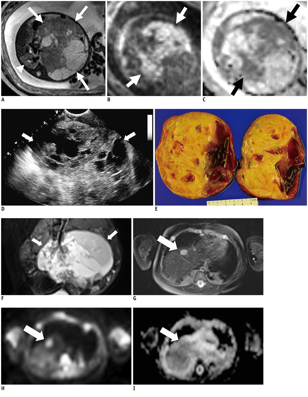
Fig. 1 Cellular mesoblastic nephroma in a female neonate. A-C. Fetal MRI shows huge mass (arrows) occupying left retroperitoneum. Solid portions show intermediate signal intensity on axial T2-weighted image (A) and right kidney (arrowhead) is visible. Solid portion of mass shows true diffusion restriction on diffusion-weighted imaging (B, white arrows) and on apparent diffusion coefficient map (C, black arrows). D. Postnatal, preoperative ultrasonography shows huge solid and cystic mass (arrows) in left abdomen. E. Gross pathology of resected mass is composed of solid and cystic portions. F-I. Whole body MRI during follow-up shows huge cystic recurrent mass (arrows) in left retroperitoneum on T2-weighted image (F) with liver dome mass (arrow), appearing as high signal on T2-weighted image (G) and diffusion restriction on diffusion-weighted imaging (H) and apparent diffusion coefficient map (I).
Reference
-
1. Bolande RP, Brough AJ, Izant RJ Jr. Congenital mesoblastic nephroma of infancy. A report of eight cases and the relationship to Wilms' tumor. Pediatrics. 1967. 40:272–278.2. Powis M. Neonatal renal tumours. Early Hum Dev. 2010. 86:607–612.3. Joshi VV, Kay S, Milsten R, Koontz WW, McWilliams NB. Congenital mesoblastic nephroma of infancy: report of a case with unusual clinical behavior. Am J Clin Pathol. 1973. 60:811–816.4. Bayindir P, Guillerman RP, Hicks MJ, Chintagumpala MM. Cellular mesoblastic nephroma (infantile renal fibrosarcoma): institutional review of the clinical, diagnostic imaging, and pathologic features of a distinctive neoplasm of infancy. Pediatr Radiol. 2009. 39:1066–1074.5. Chaudry G, Perez-Atayde AR, Ngan BY, Gundogan M, Daneman A. Imaging of congenital mesoblastic nephroma with pathological correlation. Pediatr Radiol. 2009. 39:1080–1086.6. Chan HS, Cheng MY, Mancer K, Payton D, Weitzman SS, Kotecha P, et al. Congenital mesoblastic nephroma: a clinicoradiologic study of 17 cases representing the pathologic spectrum of the disease. J Pediatr. 1987. 111:64–70.7. Puvaneswary M, Roy GT. Congenital mesoblastic nephroma: other magnetic resonance imaging findings. Australas Radiol. 1999. 43:532–534.8. Humphries PD, Sebire NJ, Siegel MJ, Olsen ØE. Tumors in pediatric patients at diffusion-weighted MR imaging: apparent diffusion coefficient and tumor cellularity. Radiology. 2007. 245:848–854.9. Zhang J, Tehrani YM, Wang L, Ishill NM, Schwartz LH, Hricak H. Renal masses: characterization with diffusion-weighted MR imaging--a preliminary experience. Radiology. 2008. 247:458–464.10. Taouli B, Thakur RK, Mannelli L, Babb JS, Kim S, Hecht EM, et al. Renal lesions: characterization with diffusion-weighted imaging versus contrast-enhanced MR imaging. Radiology. 2009. 251:398–407.11. Lowe LH, Isuani BH, Heller RM, Stein SM, Johnson JE, Navarro OM, et al. Pediatric renal masses: Wilms tumor and beyond. Radiographics. 2000. 20:1585–1603.12. Riccabona M. Imaging of renal tumours in infancy and childhood. Eur Radiol. 2003. 13:Suppl 4. L116–L129.
