Coil-Protected Embolization Technique for a Branch-Incorporated Aneurysm
- Affiliations
-
- 1Department of Radiology, The Catholic University of Korea College of Medicine, St. Vincent's Hospital, Suwon 442-723, Korea.
- 2Department of Radiology, Yonsei University College of Medicine, Severance Hospital, Seoul 120-752, Korea. bmoon21@hanmail.net
- 3Department of Radiology, Yonsei University College of Medicine, Gangnam Severance Hospital, Seoul 135-720, Korea.
- KMID: 1482796
- DOI: http://doi.org/10.3348/kjr.2013.14.2.329
Abstract
OBJECTIVE
A small branch-incorporated aneurysm is an aneurysm with a small branch incorporated into the sac or the neck. It is one of the most difficult aneurysms to treat with coil embolization. The aim of this study was to evaluate the safety and effectiveness of the coil-protected embolization technique for small-branch incorporated aneurysm.
MATERIALS AND METHODS
Fourteen aneurysms (2 ruptured and 12 unruptured) in 12 patients (mean age, 56 years, range, 40-73 years; 6 men and 6 women) were treated with the coil-protected embolization technique during the period between February 2007 and October 2011. Clinical and angiographic outcomes were retrospectively evaluated.
RESULTS
All aneurysms were successfully treated without any complications during the procedure. Immediate post-treatment angiographies demonstrated complete or near complete occlusion in 12 and incomplete occlusion in 2 patients. Two patients had a delayed small embolic infarction in the relevant posterior circulation territory and middle cerebral artery territory 10 days and 14 days later, respectively, but both recovered completely or almost completely (modified Rankin scale score [mRS score], 0 and 1, respectively). During the clinical follow-up period (mean, 21 months; range: 2-58 months), all patients reported an mRS score of 0 (n = 10) or 1 (n = 2). Vascular imaging follow-up (catheter angiography: n = 3 and MR angiography: n = 8) was available in 11 aneurysms at 6-12 months. All 11 aneurysms showed complete occlusion except for 1 minor neck recurrence that did not require further treatment.
CONCLUSION
In this series of cases, the coil-protected embolization technique seems to be feasible and effective in the treatment of small-branch incorporated aneurysms.
MeSH Terms
Figure
-
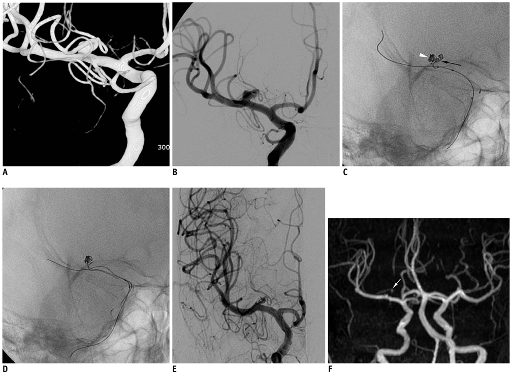
Fig. 1 Case 4. 55-year-old patient with unruptured aneurysm at right middle cerebral artery. A, B. 3D volume rendering (A) and working projection images (B) show small aneurysm with incorporated branch from which lenticulostriate arteries arises. C. While incorporated branch is protected by 3D 2/4 coil mass (black arrow) introduced through catheter facing its origin, initial coil frame (white arrowhead) is made by another coil through another microcatheter. D. After retrieval of protection coil, initial coil frame is stable. E. Post-treatment angiography shows near complete embolization of aneurysm sac with well-preserved incorporated branch. F. Six-month follow-up angiography reveals complete occlusion of aneurysm sac and preserved incorporated branch (white arrow). 3D = 3-dimensional
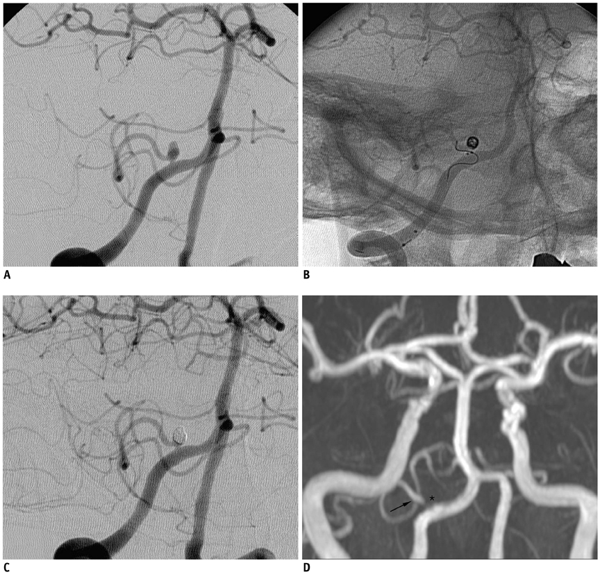
Fig. 2 Case 6. 57-year-old patient presenting with bilateral distal internal carotid artery aneurysms and right posterior inferior cerebellar artery aneurysm. A. Right vertebral angiogram in working projection shows small aneurysm with left posterior inferior cerebellar artery incorporated into aneurysm neck. B. Initial coil basket is successfully made with coil-protection technique using helical 2/4 coil. C. Final control angiogram shows complete embolization with well-preserved posterior inferior cerebellar artery. D. Six-month follow-up angiogram reveals complete occlusion of aneurysm sac (asterisk) with well-preserved right posterior inferior cerebellar artery (arrow).

Fig. 3 Case 10. 53-year-old patient presenting with unruptured left distal internal carotid artery (ICA) aneurysm and left superior cerebellar aneurysm. A. Three-dimensioal reconstruction image reveals small aneurysm with left superior cerebellar artery incorporated into aneurysm neck. B. Initial coil frame (arrowhead) is made with coil-protection (arrow) technique using helical 2/4 coil. C. After retrieval of protection coil, coil frame is stable. D. Final control angiogram reveals complete occlusion of aneurysm with preserved left superior cerebellar artery. Black asterisk indicates coil embolized left distal ICA aneurysm. E. Seven-month follow-up MR angiogram shows complete occlusion of aneurysm sac (white asterisk) and well-preserved left superior cerebellar artery.
Cited by 3 articles
-
Endovascular Coiling for a Wide-neck Bifurcated Aneurysm with Anterograde Horizontal Stenting via Microcatheter Looping: A Technical Case Report
Hyun-Jae Jeon, Jong-Hwa Park, Jong-Young Lee, Hong-Jun Jeon, Seoung-Woo Park, Byung-Moon Cho
J Cerebrovasc Endovasc Neurosurg. 2018;20(3):181-186. doi: 10.7461/jcen.2018.20.3.181.A Newly-Developed Flow Diverter (FloWise) for Internal Carotid Artery Aneurysm: Results of a Pilot Clinical Study
Byung Moon Kim, Keun Young Park, Jae Whan Lee, Joonho Chung, Dong Joon Kim, Dong Ik Kim
Korean J Radiol. 2019;20(3):505-512. doi: 10.3348/kjr.2018.0421.Parent Artery Complex Coil Protection for Side-Branched Wide-Neck Aneurysms
Keisuke Sato, Hiroshi Aoki, Shinya Jinguji, Hiroki Seto, Tsutomu Kobayashi
Neurointervention. 2022;17(2):115-120. doi: 10.5469/neuroint.2022.00136.
Reference
-
1. Kim BM, Park SI, Kim DJ, Kim DI, Suh SH, Kwon TH, et al. Endovascular coil embolization of aneurysms with a branch incorporated into the sac. AJNR Am J Neuroradiol. 2010. 31:145–151.2. Lubicz B, Lefranc F, Levivier M, Dewitte O, Pirotte B, Brotchi J, et al. Endovascular treatment of intracranial aneurysms with a branch arising from the sac. AJNR Am J Neuroradiol. 2006. 27:142–147.3. Qureshi AI, Janardhan V, Hanel RA, Lanzino G. Comparison of endovascular and surgical treatments for intracranial aneurysms: an evidence-based review. Lancet Neurol. 2007. 6:816–825.4. Kwon OK, Kim SH, Kwon BJ, Kang HS, Kim JH, Oh CW, et al. Endovascular treatment of wide-necked aneurysms by using two microcatheters: techniques and outcomes in 25 patients. AJNR Am J Neuroradiol. 2005. 26:894–900.5. Pierot L, Cognard C, Anxionnat R, Ricolfi F. CLARITY Investigators. Remodeling technique for endovascular treatment of ruptured intracranial aneurysms had a higher rate of adequate postoperative occlusion than did conventional coil embolization with comparable safety. Radiology. 2011. 258:546–553.6. Biondi A, Janardhan V, Katz JM, Salvaggio K, Riina HA, Gobin YP. Neuroform stent-assisted coil embolization of wide-neck intracranial aneurysms: strategies in stent deployment and midterm follow-up. Neurosurgery. 2007. 61:460–468. discussion 468-469.7. Kelly ME, Turner R, Gonugunta V, Woo HH, Rasmussen PA, Masaryk TJ, et al. Stent reconstruction of wide-necked aneurysms across the circle of Willis. Neurosurgery. 2007. 61:5 Suppl 2. 249–254. discussion 254-255.8. Kim BM, Kim DI, Chung EC, Kim SY, Shin YS, Park SI, et al. Endovascular coil embolization for anterior choroidal artery aneurysms. Neuroradiology. 2008. 50:251–257.9. Chae KS, Jeon P, Kim KH, Kim ST, Kim HJ, Byun HS. Endovascular coil embolization of very small intracranial aneurysms. Korean J Radiol. 2010. 11:536–541.10. Lubicz B, François O, Levivier M, Brotchi J, Balériaux D. Preliminary experience with the enterprise stent for endovascular treatment of complex intracranial aneurysms: potential advantages and limiting characteristics. Neurosurgery. 2008. 62:1063–1069. discussion 1069-1070.11. Ihn YK, Kim DI, Kim BS, Lee JM. Utility of catheter-assisted Guglielmi detachable coiling in the treatment of wide-necked aneurysms. Acta Neurochir (Wien). 2006. 148:1045–1052. discussion 1052.12. Lee JY, Seo JH, Cho YD, Kang HS, Han MH. Endovascular treatment of wide-neck intracranial aneurysms using a microcatheter protective technique: results and outcomes in 75 aneurysms. AJNR Am J Neuroradiol. 2011. 32:917–922.13. Layton KF, Cloft HJ, Gray LA, Lewis DA, Kallmes DF. Balloon-assisted coiling of intracranial aneurysms: evaluation of local thrombus formation and symptomatic thromboembolic complications. AJNR Am J Neuroradiol. 2007. 28:1172–1175.14. Pierot L, Spelle L, Leclerc X, Cognard C, Bonafé A, Moret J. Endovascular treatment of unruptured intracranial aneurysms: comparison of safety of remodeling technique and standard treatment with coils. Radiology. 2009. 251:846–855.15. Baldi S, Mounayer C, Piotin M, Spelle L, Moret J. Balloon-assisted coil placement in wide-neck bifurcation aneurysms by use of a new, compliant balloon microcatheter. AJNR Am J Neuroradiol. 2003. 24:1222–1225.16. Benitez RP, Silva MT, Klem J, Veznedaroglu E, Rosenwasser RH. Endovascular occlusion of wide-necked aneurysms with a new intracranial microstent (Neuroform) and detachable coils. Neurosurgery. 2004. 54:1359–1367. discussion 1368.17. Cottier JP, Pasco A, Gallas S, Gabrillargues J, Cognard C, Drouineau J, et al. Utility of balloon-assisted Guglielmi detachable coiling in the treatment of 49 cerebral aneurysms: a retrospective, multicenter study. AJNR Am J Neuroradiol. 2001. 22:345–351.18. Fiorella D, Albuquerque FC, Han P, McDougall CG. Preliminary experience using the Neuroform stent for the treatment of cerebral aneurysms. Neurosurgery. 2004. 54:6–16. discussion 16-17.
- Full Text Links
-
- Actions
-
Cited
- CITED
-
- Close
- Share
-
- Similar articles
-
- Retrograde Stent-assisted Coil Embolization of Wide-neck or Branch-incorporated Posterior Communicating Artery Aneurysm
- Manual Aspiration Technique to Retrieve a Prematurely Detached Coil during Cerebral Aneurysm Embolization
- Persistent Trigeminal Artery with a Cerebellar Branch and Trigeminal-Cavernous Fistula from Ruptured Aneurysm: Transarterial Coil Embolization
- Staged Y-shaped Stent Assisted Coil Embolization in a Wide-Neck Basilar Tip Aneurysm: Case Report
- Ferromagnetic Artifact Due to Metallic Embolic Fragment after Endosaccular Coil Embolization: A Case Report
