Dorsal Plating for Distal Radius Fracture
- Affiliations
-
- 1Department of Orthopedic Surgery, St Mary's Hospital, Seoul, Korea. sw.song@catholic.ac.kr
- KMID: 1480925
- DOI: http://doi.org/10.12671/jkfs.2008.21.4.334
Abstract
- No abstract available.
MeSH Terms
Figure
-
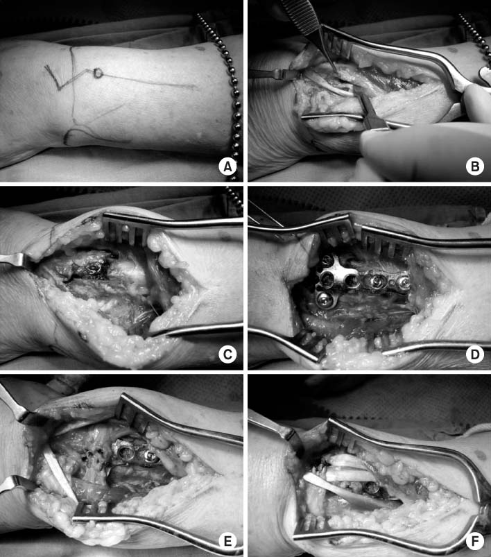
Fig. 1 (A) The zigzag midline incision over the dorsal wrist joint, centered on Lister tubercle, was designed. (B) The entire extensor retinaculum was divided into the distal and proximal halves, and the 3rd extensor compartment was opened. The 2nd and 4th extensor compartments were dissected. (C) Fracture site was reduced, (D) and temporarily fixed with K-wires. After the confirmation of proper reduction "T"-plate was fixed. (E) The half of extensor retinaculum was used to cover the transverse part of the plate, protecting the extensor tendons from the plate and screws. (F) The other half of extensor retinaculum was used to cover extensor tendon like as the original function of retinaculum.

Fig. 2 (A) After the union the patients are advised to remove the plate within 6 months to prevent the possible extensor tendon injury. Note the intact extensor tendons over the plate. (B) After the removal of screws, (C) and plate. Over and under the plate there is some thick fibrous tissue protecting the extensor tendons from injury.
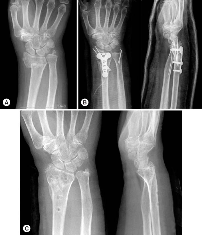
Fig. 3 (A) Left distal radius and ulnar styloid process were fractured in 74-year old female patient. (B) Dorsal plating and autogenous iliac bone graft were done for distal radius, and K-wires were inserted into the distal ulna. (C) Thirteen months after the operation the plate and screws were removed without any complications to the extensor tendons.
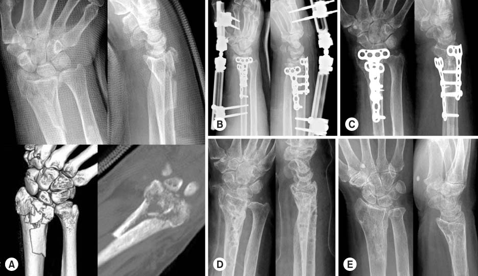
Fig. 4 (A) Sixty-nine year old female patient had broken her right wrist, with severe intraarticular fracture of distal radius. (B) Combined volar and dorsal plating were done to gather up the burst fracture fragments, and external fixation to reduce compressive pressure on the radial articular surface. (C) Seven weeks after the operation external fixator was removed. (D) Five months after the operation removal of plates and extensor tenolysis were done. (E) Twenty months after the operation. There is no evidence of post-traumatic arthritis and soft tissue complications.
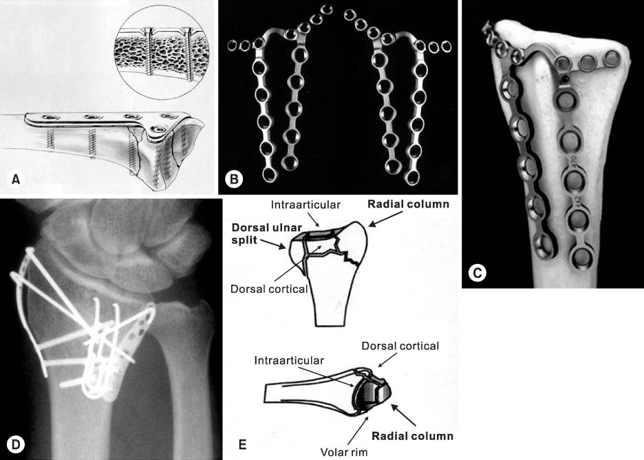
Fig. 5 Low profile plates for dorsal plating. (A) Forte plate® (Zimmer, Warsaw, IN, USA) is thinner than the conventional AO plate, and the screw head sinks into the screw hole. (B) Pi (π) plate® (Synthes, Paoli, PA) is thin and easily malleable to adapt to the complicated dorsal surface of distal radius. Left or right preference can be chosen. (C) Lister tubercle can be saved. (D) Trimed System® (Trimed, Valencia, CA) was applied to the severely comminuted distal radius fracture. (E) Trimed and 2.4-mm AO plate (Synthes, Paoli, PA) import the concept of "column" from Melone's classification of comminuted intraarticular fracture of distal radius, and give the opportunity to fix each fragment with specifically designed small plates.
Cited by 1 articles
-
Anatomical Reduction with Brick-Work Technique in Comminuted Intraarticular Distal Radius Fractures
Hyoung Min Kim, Hyung Lae Cho, Jong Woo Chae, Myung Ji Shin
J Korean Fract Soc. 2018;31(1):1-8. doi: 10.12671/jkfs.2018.31.1.1.
Reference
-
1. Axelrod TS, McMurtry RY. Open reduction and internal fixation of comminuted, intraarticular fractures of the distal radius. J Hand Surg Am. 1999; 15:1–11.
Article2. Bell JS, Wollstein R, Citron ND. Rupture of flexor pollicis longus tendon: a complication of volar plating of the distal radius. J Bone Joint Surg Br. 1998; 80:225–226.3. Carter PR, Frederick HA, Laseter GF. Open reduction and internal fixation of unstable distal radius fractures with a low-profile plate: a multicenter study of 73 fractures. J Hand Surg Am. 1998; 23:300–307.
Article4. Cross AW, Schmidt CC. Flexor tendon injuries following locked volar plating of distal radius fractures. J Hand Surg Am. 2008; 33:164–167.
Article5. Dao KD, Venn-Watson E, Shin AY. Radial artery pseudoaneurysm complication from use of AO/ASIF volar distal radius plate: a case report. J Hand Surg Am. 2001; 26:448–453.
Article6. Douthit JD. Volar plating of the dorsally comminuted fractures of the distal radius: a 6-year study. Am J Orthop. 2005; 34:140–147.7. Finsen V, Aasheim T. Initial experience with Forte plate for dorsally displaced distal radius fractures. Injury. 2000; 31:445–448.
Article8. Fitoussi F, Ip WY, Chow SP. Treatment of displaced intra-articular fractures of the distal end of the radius with plates. J Bone Joint Surg Am. 1997; 79:1303–1312.
Article9. Hahnloser D, Platz A, Amgwerd M, Trentz O. Internal fixation of distal radius fractures with dorsal dislocation: pi-plate or two 1/4 tube plates? A prospective randomized study. J Trauma. 1999; 47:760–765.
Article10. Herron M, Faraj A, Craigen MA. Dorsal plating for displaced intra-articular fractures of the distal radius. Injury. 2003; 34:497–502.
Article11. Hove LM, Nilsen PT, Furnes O, Oulie HE, Solheim E, Mölster AO. Open reduction and internal fixation of displaced intraarticular fractures of the distal radius. 31 patients followed for 3~7 years. Acta Orthop Scand. 1997; 68:59–63.
Article12. Kambouroglou GK, Axelrod TS. Complications of the AO/ASIF titanium distal radius plate system (pi plate) in internal fixation of the distal radius: a brief report. J Hand Surg Am. 1998; 23:737–741.
Article13. Keating JF, Court-Brown CM, McQueen MM. Internal fixation of volar-displaced distal radius fractures. J Bone Joint Surg Br. 1994; 76:401–405.14. Lowry KJ, Gainor BJ, Hoskins JS. Extensor tendon rupture secondary to the AO/ASIF titanium distal radius plate without associated plate failure: a case report. Am J Orthop. 2000; 29:789–791.15. Musgrave DS, Idler RS. Volar fixation of dorsally displaced distal radius fractures using 2.4-mm locking compression plates. J Hand Surg Am. 2005; 30:743–749.
Article16. Nunley JA, Rowan PR. Delayed rupture of the flexor pollicis longus tendon after inappropriate placement of the pi plate on the volar surface of the distal radius. J Hand Surg Am. 1999; 24:1279–1280.
Article17. Orbay JL. The treatment of unstable distal radius fractures with volar fixation. Hand Surg. 2000; 5:103–112.
Article18. Orbay JL, Touhami A. Current concepts in volar fixedangle fixation of unstable distal radius fractures. Clin Orthop Relat Res. 2006; 445:58–67.
Article19. Osada D, Viegas SF, Shah MA, Morris RP, Patterson RM. Comparison of different distal radius dorsal and volar fracture fixation plates: a biomechanical study. J Hand Surg Am. 2003; 28:94–104.
Article20. Peine R, Rikli DA, Hoffmann R, Duda G, Regazzoni P. Comparison of three different plating techniques for the dorsum of the distal radius: a biomechanical study. J Hand Surg Am. 2000; 25:29–33.
Article21. Ring D, Jupiter JB, Brennwald J, Büchler U, Hastings H Jr. Prospective multicenter trial of a plate for dorsal fixation of distal radius fractures. J Hand Surg Am. 1997; 22:777–784.
Article22. Schnur DP, Chang B. Extensor tendon rupture after internal fixation of a distal radius fracture using a dorsally placed AO/ASIF titanium pi plate. Arbeitsgemeinschaft für Osteosynthesefragen/Association for the Study of Internal Fixation. Ann Plast Surg. 2000; 44:564–566.
Article23. Suckel A, Spies S, Münst P. Dorsal (AO/ASIF) pi-plate osteosynthesis in the treatment of distal intraarticular radius fractures. J Hand Surg Br. 2006; 31:673–679.
Article24. Tavakolian JD, Jupoiter JB. Dorsal plating for distal radius fractures. Hand Clin. 2005; 21:341–346.
Article
- Full Text Links
-
- Actions
-
Cited
- CITED
-
- Close
- Share
-
- Similar articles
-
- Volar Plating of Distal Radius Fractures
- Dorsal Approach for Distal Radius Fractures
- Delayed Rupture of Flexor Pollicis Longus after Volar Plating for a Distal Radius Fracture
- Volar Dislocation of the Distal Radioulnar Joint Blocked by Displaced Dorsal Barton Fracture
- Complications of Distal Radius Fracture
