Pneumatization Pattern of the Frontal Recess: Relationship of the Anterior-to-Posterior Length of Frontal Isthmus and/or Frontal Recess with the Volume of Agger Nasi Cell
- Affiliations
-
- 1Department of ORL-HNS and Medical Research Institute, Pusan National University Hospital, Pusan National University School of Medicine, Busan, Korea.
- 2Department of ORL-HNS, Pusan National University Yansan Hospital, Pusan National University School of Medicine, Yangsan, Korea. rohhj@pusan.ac.kr
- KMID: 1460593
- DOI: http://doi.org/10.3342/ceo.2010.3.2.76
Abstract
OBJECTIVES
We analyzed the pneumatization pattern of the frontal recess (FR) in a Korean population. We also determined the correlation between the volume of the agger nasi cell (ANC) and the anterior-to-posterior (A-P) length of the frontal isthmus (FI) and FR.
METHODS
Multiplanar paranasal sinus computed tomography (CT) images from 105 patients who underwent endoscopic sinus surgery were reviewed. The prevalence of frontal recess cells (FRCs), thickness of the frontal beak (FB), volume of the ANC, A-P length of the FI, and FR were evaluated.
RESULTS
The ANC was identified in 96% of the patients and frontal cells (FCs) in 32% (FC type 1, 24.2%; type 2, 4.2%; type 3, 3.1%; and type 4, 0%). The prevalences of frontal bullar, suprabullar, supraorbital ethmoidal, and interfrontal sinus septal cells were 10%, 7.8%, 3.6%, and 6.8%, respectively. The A-P lengths of the FR and FI were 10.1+/-3.1 and 8.4+/-2.9 mm, respectively. The thickness of the FB was 7.8+/-1.8 mm and the volume of the ANC averaged 394.1+/-240.5 mm3. The thickness of the FB did not correlate with the volume of the ANC. In contrast, the A-P length of the FI and FR were positively correlated with the volume of the ANC.
CONCLUSION
ANCs and FCs were found in 96% and 32% of the cases in this series. FC type 4 was not seen. What appeared to be FC4 on conventional CT was identified as FBC from reconstructed parasagittal images. A large ANC increased the A-P length of the FI and FR, regardless of the thickness of the FB.
Keyword
Figure
-
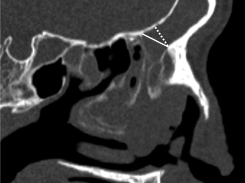
Fig. 1 A-P length of the frontal isthmus (FI; dotted line) and frontal recess (FR; solid line) on the parasagittal CT image. The anterior to posterior (A-P) length of the FI is the shortest length between the most prominent of the FB and the point of the posterior wall of the frontal sinus. The A-P length of the FR is the length between the most prominent of the FB and the superior attachment of ethmoid bullar lamella to the skull base.
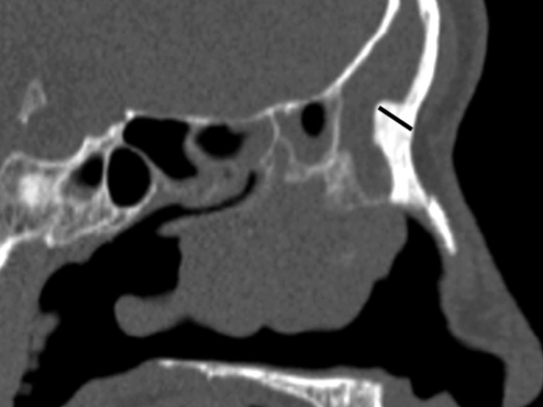
Fig. 2 Thickness of the frontal beak (FB) in prasagittal CT image. The thickness of the FB was measured on the parasagittal image where the FB showed the most prominent.
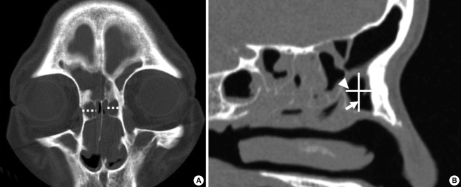
Fig. 3 Measure of the volume of the agger nasi cell (ANC) in coronal (A) and parasagittal (B) CT image. (A) The lateral diameter (dotted line) of the ANC was the longest diameter from side to side on the coronal image. (B) The longest anterior to posterior diameter (arrow head) and superior to inferior diameter (arrow) of the ANC were measured on the parasagittal image.
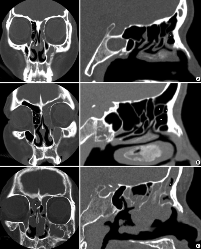
Fig. 4 Frontal cells (FCs). CT image demonstrating FC type 1, 2 and 3. Each FC cell is shown as asterisk (*) marker at A (FC type 1), B (FC type 2) and C (FC type 3).
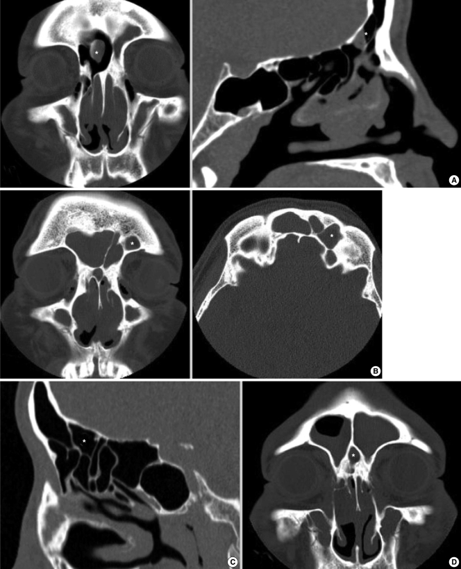
Fig. 5 CT images showing frontal bullar cell (A), supraorbital ethmoidal cell (B), suprabullar cell (C), and intersinus septal cell (D). Each cell is shown as asterisk (*) marker.

Fig. 6 Frontal bullar cell (FBC). The coronal CT image (A) shows a large frontal cell (FC) type 4 in the left frontal sinus. However, in the parasagittal CT image (B), it is the FBC, pneumatizing along the skull base into the frontal sinus from the posterior frontal recess and extending the anterior border into the frontal sinus, mistaken for the FC type 4 on coronal image.
Reference
-
1. DelGaudio JM, Hudgins PA, Venkatraman G, Beningfield A. Multiplanar computed tomographic analysis of frontal recess cells: effect on frontal isthmus size and frontal sinusitis. Arch Otolaryngol Head Neck Surg. 2005; 3. 131(3):230–235. PMID: 15781763.2. Farhat FT, Figueroa RE, Kountakis SE. Anatomic measurements for the endoscopic modified Lothrop procedure. Am J Rhinol. 2005; May–Jun. 19(3):293–296. PMID: 16011137.
Article3. Cho JH, Citardi MJ, Lee WT, Sautter NB, Lee HM, Yoon JH, et al. Comparison of frontal pneumatization patterns between Koreans and Caucasians. Otolaryngol Head Neck Surg. 2006; 11. 135(5):780–786. PMID: 17071312.
Article4. Lee JH, Lee SO. Frontal sinusitis related to anatomic variations. Korean J Otolaryngol - Head Neck Surg. 2004; 8. 47(8):751–755.5. Lee WT, Kuhn FA, Citardi MJ. 3D computed tomographic analysis of frontal recess anatomy in patients without frontal sinusitis. Otolaryngol Head Neck Surg. 2004; 9. 131(3):164–173. PMID: 15365531.
Article6. Jacobs JB, Lebowitz RA, Sorin A, Hariri S, Holliday R. Preoperative sagittal CT evaluation of the frontal recess. Am J Rhinol. 2000; Jan–Feb. 14(1):33–37. PMID: 10711330.
Article7. Kew J, Rees GL, Close D, Sdralis T, Sebben RA, Wormald PJ. Multiplanar reconstructed computed tomography images improves depiction and understanding of the anatomy of the frontal sinus and recess. Am J Rhinol. 2002; Mar–Apr. 16(2):119–123. PMID: 12030358.
Article8. Leunig A, Sommer B, Betz CS, Sommer F. Surgical anatomy of the frontal recess: is there a benefit in multiplanar CT-reconstruction? Rhinology. 2008; 9. 46(3):188–194. PMID: 18853869.9. Huang BY, Lloyd KM, DelGaudio JM, Jablonowski E, Hudgins PA. Failed endoscopic sinus surgery: spectrum of CT findings in the frontal recess. Radiographics. 2009; Jan–Feb. 29(1):177–195. PMID: 19168844.
Article10. Stammberger H, Kopp W, Dekornfeld TJ. Stammberger H, Hawke M, editors. Special endoscopic anatomy. Functional endoscopic sinus surgery: the Messerklinger technique. 1991. Philadelphia: BC Decker Publishers;p. 61–90.
- Full Text Links
-
- Actions
-
Cited
- CITED
-
- Close
- Share
-
- Similar articles
-
- Frontal Sinusitis Related to Anatomic Variations
- Effects of Frontal Recess Cells on the Development of Frontal Sinusitis
- Endoscopic Frontal Sinus Surgery
- Frontoethmoidal Cells on Computed Tomographic Analysis: The Prevalence and Relationship to Frontal Sinus/Recess Mucosal Thickening
- Anatomic Variations of the Frontal Recess and Frontal Sinusitis: Computed Tomographic Analysis
