Ciliated Foregut Cyst of the Gallbladder: A Case Report with US and CT Findings
- Affiliations
-
- 1Department of Radiology, Inha University School of Medicine, Korea. soongucho@hanmail.net
- 2Department of Internal Medicine, Inha University School of Medicine, Korea.
- 3Department of Pathology, Inha University School of Medicine, Korea.
- KMID: 1460072
- DOI: http://doi.org/10.3348/jksr.2010.62.4.373
Abstract
- A ciliated foregut cyst arising from the gall bladder is very rare with only a few reports to date. In our case, an ultrasonogram (US) showed a well-demarcated hypoechoic mass containing internal hyperechoic material in the fundus of the gallbladder. The CT scans showed a poorly enhancing round soft tissue mass in the gallbladder wall lined by an enhancing intact mucosal layer. We report here on a very rare form of the ciliated foregut cyst arising from the gall bladder including ultrasound and CT image results.
Figure
-
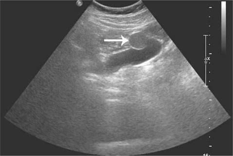
Fig. 1 An abdominal ultrasonogram shows a well-circumscribed hypoechoic mass (white arrow) with hyperechoic content in the fundus of the gallbladder.
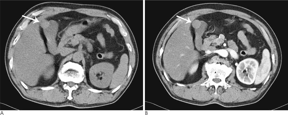
Fig. 2 A, B. The non-enhanced (A) and contrast-enhanced CT (B) scans show a well defined round mass (white arrow) with high attenuation. (Hounsfield unit: ranging from 40 to 48). The mass did not however show enhancement.
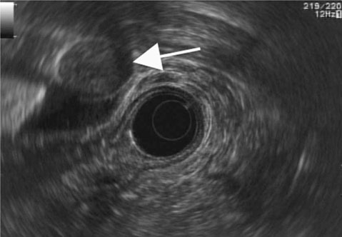
Fig. 3 An endoscopic ultrasonogram shows a well-defined hypoechoic cystic mass (white arrow) in the wall of the gallbladder.
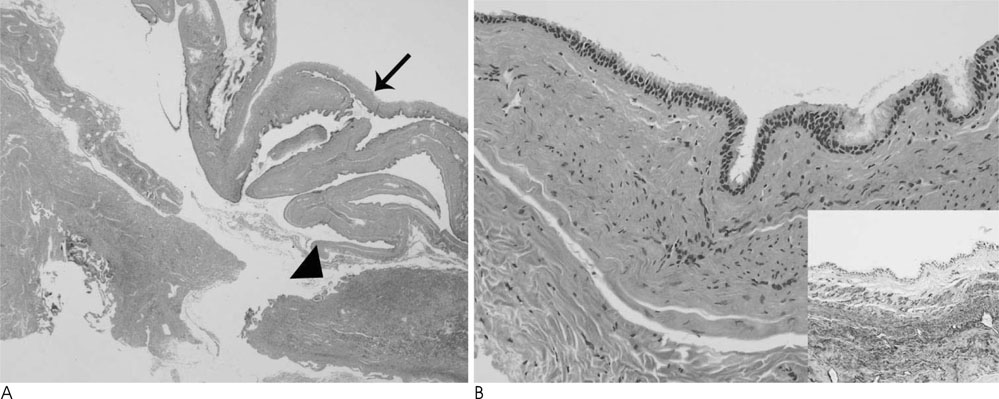
Fig. 4 A. The lower right corner shows the lumen of the gallbladder (arrowhead). The cyst (arrow) is located in the serosal area (H & E; ×12.5). B. A photomicrography shows the cyst lined with ciliated, pseudostratified columnar epithelium (H & E; ×200). Underlying smooth muscle layer that is reactive with smooth muscle actin (SMA) is visualized in the small photomicrography of left lower corner (SMA; ×200).
Reference
-
1. Muraoka A, Watanabe N, Ikeda Y, Kokudo Y, Tatemoto A, Sone Y, et al. Ciliated foregut cyst of the gallbladder: report of a case. Surg Today. 2003; 33:718–721.2. Kakitsubata Y, Kakitsubata S, Marutsuka K, Watanabe K. Epithelial cyst of the gallbladder demonstrated by ultrasonography. Radiat Med. 1995; 13:309–310.3. Benlolo D, Vilgrain V, Terris B, Zins M, Belghiti J, Menu Y. Imaging of ciliated hepatic or biliary cysts. 4 cases. Gastroenterol Clin Biol. 1996; 20:497–501.4. Nam ES, Lee HI, Kim DH, Choi CS, Kim YB, Kim JS, et al. Ciliated foregut cyst of the gallbladder: a case report and review of the literature. Pathol Int. 2000; 50:427–430.5. Hirono S, Tanimura H, Yokoyama S, Uchiyama K, Tani M, Onishi H, et al. Clinical features of ciliated foregut cyst of the gallbladder: a rare entity of cystic lesion in the gallbladder. Dig Dis Sci. 2002; 47:1817–1820.6. Jacobs E, Ardichvili D, D'avanzo E, Penneman R, Van Gansbeke D. Cyst of the gallbladder. Dig Dis Sci. 1991; 36:1796–1802.7. Cureton RJ, Newcomb JF. Multilocular cyst of the gallbladder. Br J Surg. 1961; 48:577–580.8. Kadoya M, Matsui O, Nakanuma Y, Yoshikawa J, Arai K, Takashima T, et al. Ciliated hepatic foregut cyst: radiologic features. Radiology. 1990; 175:475–477.9. Kimura A, Makuuchi M, Takayasu K, Sakamoto M, Hirohashi S. Ciliated hepatic foregut cyst with solid tumor appearance on CT. J Comput Assist Tomogr. 1990; 14:1016–1018.
- Full Text Links
-
- Actions
-
Cited
- CITED
-
- Close
- Share
-
- Similar articles
-
- Ciliated foregut cyst of the gallbladder: a case report and literature review
- Ciliated Foregut Cyst of the Liver: Report of a case
- A Case of Ciliated Foregut Cyst of the Gallbladder
- Ciliated Foregut Cyst and Accessory Spleen in the Pancreas: A Case Report and Literature Review
- A case of ciliated hepatic foregut cyst treated by laparoscopic excision
