Korean Circ J.
2010 Sep;40(9):468-470. 10.4070/kcj.2010.40.9.468.
Long Journey of Sclerosant From the Esophagus to the Right Atrium
- Affiliations
-
- 1Department of Cardiology, Ajou University School of Medicine, Suwon, Korea. shinjh@ajou.ac.kr
- 2Department of Pulmonary and Critical Care Medicine, Ajou University School of Medicine, Suwon, Korea.
- KMID: 1456141
- DOI: http://doi.org/10.4070/kcj.2010.40.9.468
Abstract
- A 34-year-old man, who had been treated with an endoscopic injection of a mixture of n-butyl-2-cyanoacrylate (Histoacryl) and Lipiodol for control of variceal bleeding 6 months previously, presented with an intracardiac mass in the right atrium (RA). Two-dimensional echocardiography revealed an intracardiac mass in the RA that appeared to extend from the inferior vena cava. The origin of the sclerosant was traced by computed tomography (CT). This is a very rare case in which the sclerosant migration route was demonstrated by CT scan. The findings of this case suggest that the systemic migration of sclerosant into an intracardiac chamber should be considered in patients with an intracardiac mass, especially with a history of previous sclerotherapy for variceal bleeding.
MeSH Terms
Figure
-
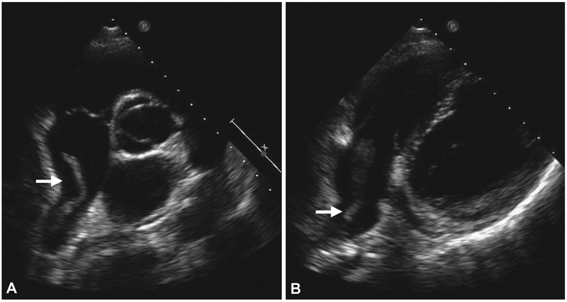
Fig. 1 Transthoracic echocardiography shows an elongated intracardiac mass in the right atrium (arrow), which appears to extend from the inferior vena cava. A: parasternal short axis view, aortic valve level. B: off-axis modified four-chamber view.

Fig. 2 Portal-phase abdominal computed tomography scans with coronal reconstruction demonstrate a partial radio-opaque and radiolucent intraluminal material consistent with a mixture of sclerosant and thrombus, which originates from the gastroesophageal junction (1) and forms an intraluminal and intracardiac mass traversing a gastrorenal shunt (2), the left renal vein (3), the inferior vena cava (4) and right atrium (5).
Reference
-
1. See A, Florent C, Lamy F, Levy VG, Bouvry M. Cerebrovascular accident after endoscopic obturation of esophageal varices with isobutyl-2-cyanoacylate in 2 patients. Gastroenterol Clin Biol. 1986. 10:604–607.2. Thakeb F, Kader S, Salama Z, Abdel Raouf T, Abdel Kader S, Abdel Hamid H. The value of the combined use of n-butyl-2-cyanoacrylate and ethanolamine oleate in the management of bleeding esophagogastric varices. Endoscopy. 1995. 27:358–364.3. Moustafa I, Omar MM, Nooh A. Endoscopic control of gastric variceal bleeding with butyl cyanoacrylate in patients with schisiosomiasis. J Egypt Soc Parasitol. 1997. 27:405–410.4. Huang YH, Yeh HZ, Chen GH, et al. Endoscopic treatment of bleeding gastric varices by N-butyl-2-cyanoacrylate (Histoacryl) injection: long-term efficacy and safety. Gastrointest Endosc. 2000. 52:160–167.5. Anderson JM, Gibbons DF. The new generation of biochemical polymers. Biomater Med Devices Artif Organs. 1974. 2:235–248.6. D'Imperio N, Piemontese A, Baroncini D, et al. Evaluation of undiluted N-butyl-2-cyanoacrylate in the endoscopic treatment of upper gastrointestinal tract varices. Endoscopy. 1996. 28:239–243.7. Dhiman RK, Chawla Y, Taneja S, Biswas R, Sharma TR, Dilawari JB. Endoscopic sclerotherapy of gastric variceal bleeding with N-butyl-2-cyanoacrylate. J Clin Gastroenterol. 2002. 35:222–227.8. Cheng PN, Sheu BS, Chen CY, Chang TT, Lin XZ. Splenic infarction after Histoacryl injection for bleeding gastric varices. Gastrointest Endosc. 1998. 48:426–427.9. Hwang SS, Kim HH, Park SH, et al. N-butyl-2-cyanoacrylate pulmonary embolism after endoscopic injection sclerotherapy for gastric variceal bleeding. J Comput Assist Tomogr. 2001. 25:16–22.10. Javed A, Salamat A. N-butyl-2-cyanoacrylate and lipiodol pulmonary embolism (glue embolism). J Ayub Med Coll Abbottabad. 2008. 20:143–145.11. Lee SE, Chang SA, Kim MS, et al. Acrylic cement foreign body and thrombus in right atrium causing pulmonary embolism after percutaneous vertebroplasty. Korean Circ J. 2006. 36:713–715.
- Full Text Links
-
- Actions
-
Cited
- CITED
-
- Close
- Share
-
- Similar articles
-
- Automatic Sclerosant Injection Technique of Mechanochemical Ablation with ClariVein Using a Syringe Pump for the Treatment of Varicose Veins
- A Case of Long-Segment Barrett's Esophagus with Mixed Connective Tissue Disease
- A Case of Recurrence after Endoscopic Submucosal Dissection of Esophageal Adenocarcinoma Arising from Barrett's Esophagus
- Study for Mongyu Dowondo ‒ Dream Journey to the Peach Blossom Land ‒ in the Perspective of the Unconscious: Centering on Dream Interpretation of Prince An Pyeong
- Psychoanalytic Perspectives of the Development of an Adolescent Through Loss and Mourning in a Novel, ‘The Neverending Story’
