Determination of midsagittal plane for evaluation of facial asymmetry using three-dimensional computed tomography
- Affiliations
-
- 1Department of Oral and Maxillofacial Radiology and Dental Research Institute, School of Dentistry, Seoul National University, Seoul, Korea. future3@snu.ac.kr
- 2Department of Oral and Maxillofacial Surgery, Ilsan Paik Hospital, Inje University, Goyang, Korea.
- 3Department of Oral and Maxillofacial Surgery, Seoul National University Dental Hospital, Seoul, Korea.
- 4Section of Orthodontics, Samsung Medical Center, Seoul, Korea.
- KMID: 1449939
- DOI: http://doi.org/10.5624/isd.2011.41.2.79
Abstract
- PURPOSE
The aim of the present study was to investigate the disagreement of cephalometric analysis depending on the reference determination of midsagittal plane on three-dimensional computed tomography.
MATERIALS AND METHODS
A total of 102 young women with class III dentofacial deformity were evaluated using three-dimensional computed tomography. The cranial and facial midsagittal planes were defined and the amounts of jaw deviation were calculated. The amounts of jaw deviation were compared with paired t-test (2-tailed) and Bland-Altman plot was drawn.
RESULTS
The landmark tracing were reproducible (r> or =.978). The jaws relative to the cranial midsagittal plane were 10-17 times more significantly deviated than to the facial midsagittal plane (P<.001). Bland-Altman plot demonstrated that the differences between the amounts of jaw deviation from two midsagittal planes were not normally distributed versus the average of the amounts of jaw deviation from two midsagittal planes.
CONCLUSION
The cephalometric analyses of facial asymmetry were significantly inconsistent depending on the reference determination of midsagittal plane. The reference for midsagittal plane should be carefully determined in three-dimensional cephalometric analysis of facial asymmetry of patients with class III dentofacial deformity.
MeSH Terms
Figure
-
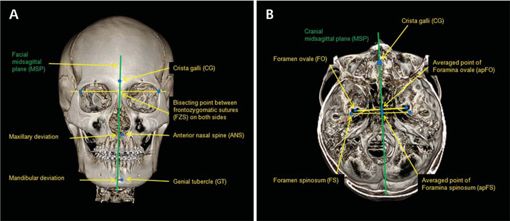
Fig. 1 Landmarks and jaw deviation. A. The jaw deviation is determined as a perpendicular distance between midpoint (ANS and GT) and midsagittal plane (MSP), B. The averaged points of foramina ovale (FO) and foramina spinosum (FS) on both sides are depicted.
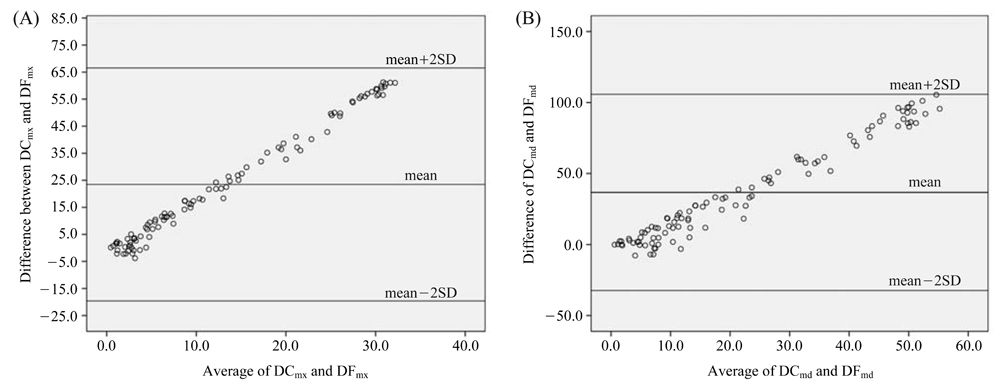
Fig. 2 Bland-Altman plot for jaw deviation to two midsagittal planes (MSP). The x-axis indicates the average (mm) of DCs and DFs, whereas the y-axis indicates the difference (mm) between DCs and DFs. The limits of agreement (mean difference±2 SD) are graphed by the horizontal lines. A. For the maxillary deviation, B. For the mandibular deviation. Both plots demonstrate the DCs and DFs are not normally distributed by the Bland-Altman method. This means DCs are larger than DFs as the jaw midpoints (ANS and GT) are distant from the MSP.
Cited by 3 articles
-
Deviation of landmarks in accordance with methods of establishing reference planes in three-dimensional facial CT evaluation
Kaeng Won Yoon, Suk-Ja Yoon, Byung-Cheol Kang, Young-Hee Kim, Min Suk Kook, Jae-Seo Lee, Juan Martin Palomo
Imaging Sci Dent. 2014;44(3):207-212. doi: 10.5624/isd.2014.44.3.207.A comparative study of the deviation of the menton on posteroanterior cephalograms and three-dimensional computed tomography
Hee Jin Lee, Sungeun Lee, Eun Joo Lee, In Ja Song, Byung-Cheol Kang, Jae-Seo Lee, Hoi-Jeong Lim, Suk-Ja Yoon
Imaging Sci Dent. 2016;46(1):33-38. doi: 10.5624/isd.2016.46.1.33.The location of midfacial landmarks according to the method of establishing the midsagittal reference plane in three-dimensional computed tomography analysis of facial asymmetry
Min Sun Kim, Eun Joo Lee, In Ja Song, Jae-Seo Lee, Byung-Cheol Kang, Suk-Ja Yoon
Imaging Sci Dent. 2015;45(4):227-232. doi: 10.5624/isd.2015.45.4.227.
Reference
-
1. Finlay LM. Craniometry and cephalometry: a history prior to the advent of radiography. Angle Orthod. 1980. 50:312–321.2. The American Heritage medical dictionary. 2008. Boston: Houghton Mifflin.3. Trpkova B, Prasad NG, Lam EW, Raboud D, Glover KE, Major PW. Assessment of facial asymmetries from posteroanterior cephalograms: validity of reference lines. Am J Orthod Dentofacial Orthop. 2003. 123:512–520.
Article4. Suri S, Utreja A, Khandelwal N, Mago SK. Craniofacial computerized tomography analysis of the midface of patients with repaired complete unilateral cleft lip and palate. Am J Orthod Dentofacial Orthop. 2008. 134:418–429.
Article5. Lagravere MO, Hansen L, Harzer W, Major PW. Plane orientation for standardization in 3-dimensional cephalometric analysis with computerized tomography imaging. Am J Orthod Dentofacial Orthop. 2006. 129:601–604.6. Lagravere MO, Major PW. Proposed reference point for 3-dimensional cephalometric analysis with cone-beam computerized tomography. Am J Orthod Dentofacial Orthop. 2005. 128:657–660.7. Olszewski R, Cosnard G, Macq B, Mahy P, Reychler H. 3D CT-based cephalometric analysis: 3D cephalometric theoretical concept and software. Neuroradiology. 2006. 48:853–862.
Article8. Deguchi T Sr, Katashiba S, Inami T, Foong KW, Huak CY. Morphologic quantification of the maxilla and the mandible with cone-beam computed tomography. Am J Orthod Dentofacial Orthop. 2010. 137:218–222.
Article9. Muramatsu A, Nawa H, Kimura M, Yoshida K, Maeda M, Katsumata A, et al. Reproducibility of maxillofacial anatomic landmarks on 3-dimensional computed tomographic images determined with the 95% confidence ellipse method. Angle Orthod. 2008. 78:396–402.
Article10. Fleiss JL, Chilton NW. The measurement of interexaminer agreement on periodontal disease. J Periodontal Res. 1983. 18:601–606.
Article11. Bland JM, Altman DG. Statistical methods for assessing agreement between two methods of clinical measurement. Lancet. 1986. 1:307–310.
Article12. Adams GL, Gansky SA, Miller AJ, Harrell WE Jr, Hatcher DC. Comparison between traditional 2-dimensional cephalometry and a 3-dimensional approach on human dry skulls. Am J Orthod Dentofacial Orthop. 2004. 126:397–409.
Article13. Pirttiniemi PM. Associations of mandibular and facial asymmetries - a review. Am J Orthod Dentofacial Orthop. 1994. 106:191–200.14. Galaburda AM, LeMay M, Kemper TL, Geschwind N. Right-left asymmetrics in the brain. Science. 1978. 199:852–856.15. Severt TR, Proffit WR. The prevalence of facial asymmetry in the dentofacial deformities population at the University of North Carolina. Int J Adult Orthodon Orthognath Surg. 1997. 12:171–176.16. Haraguchi S, Takada K, Yasuda Y. Facial asymmetry in subjects with skeletal Class III deformity. Angle Orthod. 2002. 72:28–35.17. Baek SH, Cho IS, Chang YI, Kim MJ. Skeletodental factors affecting chin point deviation in female patients with class III malocclusion and facial asymmetry: a three-dimensional analysis using computed tomography. Oral Surg Oral Med Oral Pathol Oral Radiol Endod. 2007. 104:628–639.
Article18. Proffit WR, Phillips C, Dann C 4th. Who seeks surgical-orthodontic treatment? Int J Adult Orthodon Orthognath Surg. 1990. 5:153–160.19. Enlow DH, McNamara JA Jr. The neurocranial basis for facial form and pattern. Angle Orthod. 1973. 43:256–270.20. Friede H. Normal development and growth of the human neurocranium and cranial base. Scand J Plast Reconstr Surg. 1981. 15:163–169.
Article21. Janson G, de Lima KJ, Woodside DG, Metaxas A, de Freitas MR, Henriques JF. Class II subdivision malocclusion types and evaluation of their asymmetries. Am J Orthod Dentofacial Orthop. 2007. 131:57–66.
Article22. Kanomi R, Hidaka O, Yamada C, Takada K. Asymmetry in the condylar long axis and first molar rotation. J Dent Res. 2004. 83:109–114.23. Williamson PC, Major PW, Nebbe B, Glover KE. Landmark identification error in submentovertex cephalometrics. A computerized method for determining the condylar long axis. Oral Surg Oral Med Oral Pathol Oral Radiol Endod. 1998. 86:360–369.24. Lascala CA, Panella J, Marques MM. Analysis of the accuracy of linear measurements obtained by cone beam computed tomography (CBCT-NewTom). Dentomaxillofac Radiol. 2004. 33:291–294.
Article25. Waitzman AA, Posnick JC, Armstrong DC, Pron GE. Craniofacial skeletal measurements based on computed tomography: Part II. Normal values and growth trends. Cleft Palate Craniofac J. 1992. 29:118–128.
Article26. Sgouros S, Natarajan K, Hockley AD, Goldin JH, Wake M. Skull base growth in childhood. Pediatr Neurosurg. 1999. 31:259–268.
Article27. Enlow DH, Hans MG. Essentials of facial growth. 1996. Philadelphia: WB Saunders.28. Lee KH, Hwang HS, Curry S, Boyd RL, Norris K, Baumrind S. Effect of cephalometer misalignment on calculations of facial asymmetry. Am J Orthod Dentofacial Orthop. 2007. 132:15–27.
Article
- Full Text Links
-
- Actions
-
Cited
- CITED
-
- Close
- Share
-
- Similar articles
-
- Comparison of midsagittal reference plane in PA cephalogram and 3D CT
- Deviation of landmarks in accordance with methods of establishing reference planes in three-dimensional facial CT evaluation
- Differences in positions of cone-beam computed tomography landmarks in patients with skeletal Class III facial asymmetry according to midsagittal planes
- The location of midfacial landmarks according to the method of establishing the midsagittal reference plane in three-dimensional computed tomography analysis of facial asymmetry
- Three dimensional CT analysis of facial asymmetry
