J Korean Assoc Oral Maxillofac Surg.
2011 Aug;37(4):264-271. 10.5125/jkaoms.2011.37.4.264.
A comparative study on the change of postoperative facial hard tissue profile after maxillary rotational surgery
- Affiliations
-
- 1Department of Oral and Maxillofacial Surgery, School of Dentistry, Pusan National University, Yangsan, Korea. inkchung@pusan.ac.kr
- KMID: 1449058
- DOI: http://doi.org/10.5125/jkaoms.2011.37.4.264
Abstract
- PURPOSE
This study evaluated retrospectively the postsurgical facial hard tissue profile of a Le Fort I osteotomy with/without posterior impaction and rigid internal fixation to correct mandibular prognathism. After observing a difference between the two groups, this measurement was used to prepare a treatment plan for 2-jaw surgery. Patients and Methods: Thirty patients who had undergone orthognathic surgery in Pusan National University Dental Hospital were enrolled in this study. Fifteen patients were treated using a Le Fort I osteotomy with posterior impaction and mandibular setback bilateral sagittal split ramus osteotomy, and the other fifteen patients were treated without posterior impaction. The preoperative (T0), immediate postoperative (T1) and six-month follow-up period (T2) cephalograms were taken and difference between T1-T0 and T2-T2 was analyzed.
RESULTS
Both groups was FH-ABp, SNB and ANB showed significant changes in the measurement, whereas only the posterior impaction group showed a change in the SN-U1, occlusal plane, posterior facial height, surgical movement difference from the L1 and B-point. There was no significant statistical change between the immediate postoperative (T1) and six-month follow-up (T2) hard tissue analysis in the two groups.
CONCLUSION
A Le Fort I osteotomy with posterior impaction is considerable for patients with a flat occlusal plane angle, large posterior facial height, prominent B-point, pogonion and labioversed incisal inclination if the indications are well chosen.
MeSH Terms
Figure
-
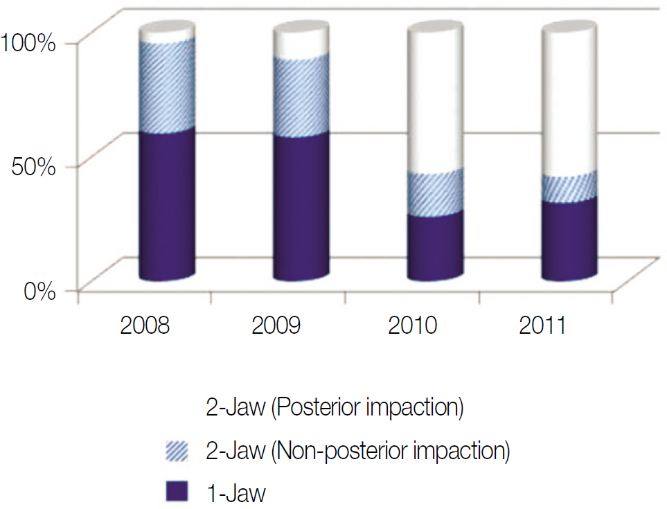
Fig. 1. Annual trend of the surgical treatment modality of orthognathic surgery since 2008 in Department of Oral and Maxillofacial Sugery, Pusan National University Dental Hospital.

Fig. 2. Anatomic landmark used in this study.

Fig. 3. Reference line.
Reference
-
1. Enacar A, Taner T, Manav O. Effects of single- or double-jaw surgery on vertical dimension in skeletal Class III patients. Int J Adult Orthodon Orthognath Surg. 2001; 16:30–5.2. Reyneke JP, Evans WG. Surgical manipulation of the occlusal plane. Int J Adult Orthodon Orthognath Surg. 1990; 5:99–110.3. Wolford LM, Chemello PD, Hilliard F. Occlusal plane alteration in orthognathic surgery-Part I: Effects on function and esthetics. Am J Orthod Dentofacial Orthop. 1994; 106:304–16.
Article4. Praveen K, Narayanan V, Muthusekhar MR, Baig MF. Hypotensive anesthesia and blood loss in orthognathic surgery: a clinical study. Br J Oral Maxillofac Surg, 2001:39;138-40.5. Chang HH, Ryu SH, Kang JH, Lee SH, Kim JS. Blood loss and hematologic change after orthognathic surgery. J Korean Oral Maxillofac Surg. 2001; 27:435–41.6. Reyneke JP, Bryant RS, Suuronen R, Becher PJ. Postoperative skeletal stability following clockwise and counter-clockwise rotation of the maxillomandibular complex compared to conventional orthognathic treatment. Br J Oral Maxillofac Surg. 2007; 45:56–64.
Article7. Wolford LM, Chemello PD, Hilliard F. Occlusal plane alteration in orthognathic surgery-Part I: Effects on function and esthetics. Am J Orthod Dentofacial Orthop. 1994; 106:304–16.
Article8. Travess HC, Newton JT, Sandy JR, Williams AC. The development of a patient-centered measure of the process and outcome of combined orthodontic and orthognathic treatment. J Orthod. 2004; 31:220–34.
Article9. Cunningham SJ, Garratt AM, Hunt NP. Development of a condition-specific quality of life measure for patients with dentofacial deformity: II. Validity and responsiveness testing. Community Dent Oral Epidemiol. 2002; 30:81–90.
Article10. Burke L, Croucher R. Criteria of good dental practice generated by general dental practitioners and patients. Int Dent J. 1996; 46:3–9.11. Laufer D, Glick D, Gutman D, Sharon A. Patients motivation and response to surgical correction of prognathism. Oral surg Oral Med Oral Pathol. 1976; 41:309–13.12. Jeong MH, Choi JH, Kim BH, Kim SG, Nahm DS. Soft tissue changes after double jaw rotation surgery in skeletal class III malocclusion. J Korean Oral Maxillofac Surg. 2006; 32:559–65.13. Epker BN, Schendel SA. Total maxillary surgery. Int J Oral Surg. 1980; 9:1–24.
Article14. LaBanc JP, Epker BN. Changes of the hyoid bone and tongue following advancement of the mandible. Oral Surg Oral Med Oral Pathol. 1984; 57:351–6.
Article15. Kim BH, et al. Treatment goals and planning in class III 2-jaw surgery-the contribution of jaw rotation. J Korean Foundation for Gnatho-orthodontic research. 2005; 7:39–51.16. Yang SD, Suhr CH. F-H to AB plane angle(FABA) for assessment of anteroposterior jaw relationships. Angle Orthod. 1995; 65:223–31.17. Kim KH, Choy KC, Kim HG, Park KH. Cephalometric Norms of the hard tissues of Korean orthognathic surgery. J Korean Oral Maxillofac Surg. 2001; 27:221–30.18. Holdaway RA. A soft-tissue cephalometric analysis and its use in orthodontic treatment planning. Part I. Am J Orthod. 1983; 84:1–28.
Article
- Full Text Links
-
- Actions
-
Cited
- CITED
-
- Close
- Share
-
- Similar articles
-
- A cephalometric study on the changes of soft tissue profile (upper lip and nose) following two-jaw surgery
- Soft Hard Tissue Changes Following Anterior Segmental Sorcery In Bimaxillary Protrusion
- A STUDY ON CHANGE OF THE SOFT TISSUE FACIAL PROFILE AFTER ORTHOGNATHIC SORCERY IN PATIENTS WITH THE MANDIBULAR PROGNATHISM
- Prediction of frontal soft tissue changes after mandibular surgery in facial asymmetry individuals
- Stability And Soft Tissue Changes Following Advancement Le Fort I Osteotomy In The Cleft Lip And Palate Patients
