Abnormal patterns of the renal veins
- Affiliations
-
- 1Department of Anatomical Sciences, School of Medicine, Kurdistan University of Medical Sciences, Sanandaj, Iran. hadi_anjam@yahoo.com
- 2Department of Anatomical Sciences, School of Medicine, Shiraz University of Medical Sciences, Shiraz, Iran.
- 3Department of Urology, Moradi Hospital, School of Medicine, Rafsanjan University of Medical Sciences, Rafsanjan, Iran.
- KMID: 1447454
- DOI: http://doi.org/10.5115/acb.2012.45.1.57
Abstract
- Knowledge of the renal vascular anatomy may greatly contribute to the success of surgical, invasive and radiological procedures of the retroperitoneal region. Here, morphometric and histological studies of a human cadaveric specimen presented a complex, anomalous pattern of renal veins. The left renal vein had an oblique retro-aortic course and received two lumbar veins. It bifurcated near its drainage point into the inferior vena cava. The right renal vein received the right testicular vein. In addition, the left kidney was located at a low position. The spleen was enlarged. The present case is unique and provides information that may help surgeons or angiologists to apply safer interventions.
Keyword
MeSH Terms
Figure
-
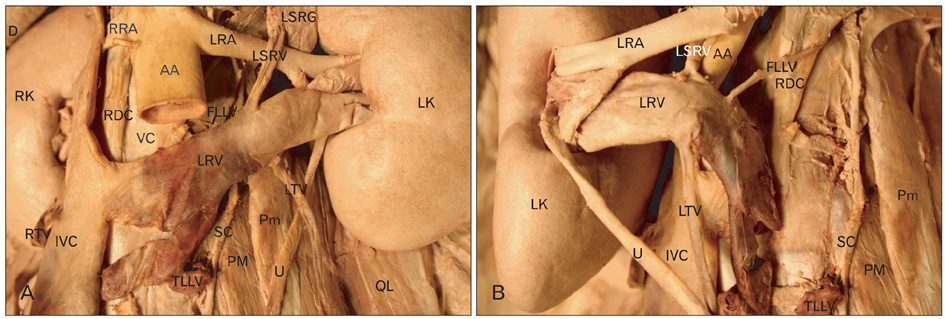
Fig. 1 Left retroperitoneal space dissection shows the abnormal drainage of a retro-aortic bifid left renal vein into the inferior vena cava. The variant termination of the 1st and 3rd lumbar veins into the left renal vein is also seen. (A) The abdominal aorta is dissected to show the bifid termination of the retro-aortic left renal vein. (B) The left kidney and structures that pass through its hilum are shown in their reflected position. AA, abdominal aorta; D, diaphragm; FLLV, first left lumbar vein; IVC, inferior vena cava; LK, left kidney; LRA, left renal artery; LRV, left renal vein; LSRG, left suprarenal gland; LSRV, left suprarenal vein; LTV, left testicular vein; PM, psoas major; Pm, psoas minor; QL, quadrates lumborum; RDC, right diaphragmatic crus; RK, right kidney; RRA, right renal artery; RTV, right testicular vein; SC, sympathetic chain; TLLV, third left lumbar vein; U, ureter; VC, vertebral column.
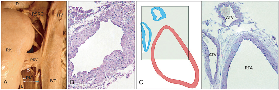
Fig. 2 Right retroperitoneal space dissection shows; (A) the abnormal drainage of additional testicular vein into the right renal vein. (B) Light micrograph illustrating a histological section of testicular vein before drainage into the right renal vein. (C) Right testicular artery with accompanying veins in a common sheath (right, schematic illustration; left, histological section) (B, ×200; C, ×100). ATV, additional testicular vein; D, diaphragm; HV, hepatic vein; IVC, inferior vena cava; PM, psoas major; RK, right kidney; RRV, right renal vein; RSRG, right suprarenal gland; RTA, right testicular artery; U, ureter.

Fig. 3 Retroperitoneal space dissection. (A) The greatly enlarged spleen forced the left kidney below the level of the right kidney. (B) The isolated enlarged spleen. LK, left kidney; RK, right kidney; S, spleen; SA, splenic artery; SV, splenic vein.
Cited by 1 articles
-
Multiple renal veins clogging the hilum of the right kidney
Satheesha B Nayak, Narendra Pamidi, Vasanthakumar Packirisamy, Soumya Kodimajalu Vasudeva
Anat Cell Biol. 2023;56(1):141-144. doi: 10.5115/acb.22.109.
Reference
-
1. Satyapal KS, Kalideen JM, Haffejee AA, Singh B, Robbs JV. Left renal vein variations. Surg Radiol Anat. 1999. 21:77–81.2. Senecail B, Bobeuf J, Forlodou P, Nonent M. Two rare anomalies of the left renal vein. Surg Radiol Anat. 2003. 25:465–467.3. Malcic-Gürbüz J, Akalin A, Gümüşcü B, Cavdar S. Clinical implications of concomitant variations of the testicular, suprarenal and renal veins: a case report. Ann Anat. 2002. 184:35–39.4. Benedetti E, Troppmann C, Gillingham K, Sutherland DE, Payne WD, Dunn DL, Matas AJ, Najarian JS, Grussner RW. Short- and long-term outcomes of kidney transplants with multiple renal arteries. Ann Surg. 1995. 221:406–414.5. Jetti R, Jevoor P, Vollala VR, Potu BK, Ravishankar M, Virupaxi R. Multiple variations of the urogenital vascular system in a single cadaver: a case report. Cases J. 2008. 1:344.6. Kumar S, Neyaz Z, Gupta A. The utility of 64 channel multidetector CT angiography for evaluating the renal vascular anatomy and possible variations: a pictorial essay. Korean J Radiol. 2010. 11:346–354.7. Bass JE, Redwine MD, Kramer LA, Huynh PT, Harris JH Jr. Spectrum of congenital anomalies of the inferior vena cava: cross-sectional imaging findings. Radiographics. 2000. 20:639–652.8. Field S, Saxton H. Venous anomalies complicating left adrenal catheterization. Br J Radiol. 1974. 47:219–225.9. Macchi V, Parenti A, De Caro R. Pivotal role of the sub-supracardinal anastomosis in the development and course of the left renal vein. Clin Anat. 2003. 16:358–361.10. Arslan H, Etlik O, Ceylan K, Temizoz O, Harman M, Kavan M. Incidence of retro-aortic left renal vein and its relationship with varicocele. Eur Radiol. 2005. 15:1717–1720.11. Holden A, Smith A, Dukes P, Pilmore H, Yasutomi M. Assessment of 100 live potential renal donors for laparoscopic nephrectomy with multi-detector row helical CT. Radiology. 2005. 237:973–980.
- Full Text Links
-
- Actions
-
Cited
- CITED
-
- Close
- Share
-
- Similar articles
-
- Normal variations of renal vessels based upon the study of 240 living-donor nephrectomies
- Case Report on Horseshoe Kidney
- Angiographic analysis of renal artery and vein in 85 candidates of renal transplant dornor
- Common Variations of Renal Vessels in Donor Kidneys
- Multi-Detector CT Findings of Double Retroaortic Left Renal Veins
