Primary leiomyosarcoma of the pancreas
- Affiliations
-
- 1Division of Hepato-Biliary-Pancreatic Surgery, Department of Surgery, Chonnam National University Medical School, Gwangju, Korea. ckcho@jnu.ac.kr
- 2Department of Radiology, Chonnam National University Medical School, Gwangju, Korea.
- 3Department of Pathology, Chonnam National University Medical School, Gwangju, Korea.
- KMID: 1445790
- DOI: http://doi.org/10.4174/jkss.2011.81.Suppl1.S69
Abstract
- Primary sarcomas of the pancreas are extremely rare, accounting for 0.1% of malignant pancreatic (non-islet) neoplasms. Pancreatic leiomyosarcoma is a highly aggressive malignancy that spreads in a similar manner to gastric leiomyosarcoma, i.e., by adjacent organ invasion, hematogenous spread, and lymph node metastasis. These tumors are large at the time of diagnosis and are usually found at an advanced stage. We report a case of a 70-year-old female with intermittent right upper quadrant abdominal discomfort. Radiological, histopathological, and immunohistochemical studies revealed the tumor to be a primary leiomyosarcoma of the pancreas. Herein, we describe a patient with a primary leiomyosarcoma of the pancreas who presented with clinical and radiological findings indicative of a mass in the pancreatic head.
Keyword
MeSH Terms
Figure
-
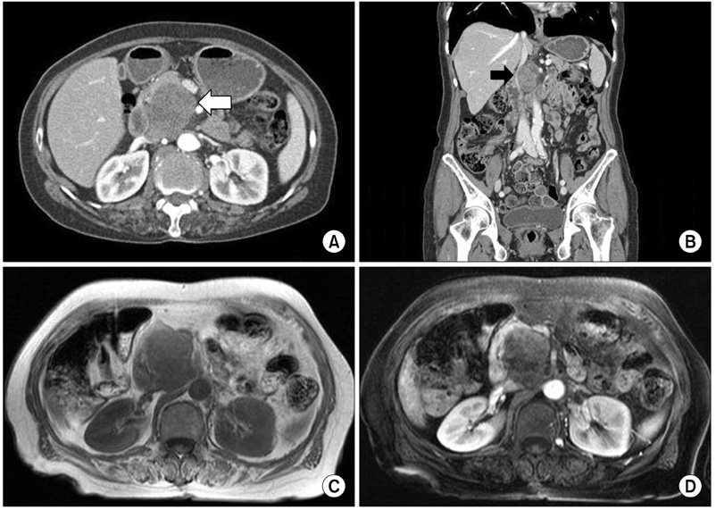
Fig. 1 Abdominal computed tomography (CT) scan shows growth of the tumor, which is heterogeneously enhanced by contrast media (A, white arrow). CT scan shows the tumor, which is abutting to the inferior vena cava (B, black arrow). Magnetic resonance imaging shows a 5.3 × 4 cm heterogenously enhanced mass (C, D).
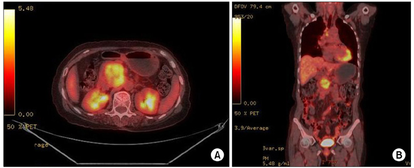
Fig. 2 Whole body [18F]-fluoro-deoxy glucose positron emission tomography/computed tomography demonstrates a 5.6 × 3.8 cm size hypermetabolic mass arising from the pancreatic head in the retropancreatic space.

Fig. 3 The gross pathological examination reveals a 5 × 5 cm multiseptated mass in the pancreatic head. The cut surface of the tumor is whitish and shows signs of internal hemorrhage and partial myxoid change (A). Interface of tumor (left) and compressed adjacent pancreatic tissue with fibrosis (B, H&E, ×40). The mass contains interlacing spindle-shaped cells with varying degrees of pleomorphism and atypia (C, H&E, ×200). The immunohistochemical findings show strong immunoreactivity of the tumor cells to actin (D, labeled streptavidin-biotin, original magnification ×200).
Reference
-
1. Baylor SM, Berg JW. Cross-classification and survival characteristics of 5,000 cases of cancer of the pancreas. J Surg Oncol. 1973. 5:335–358.2. Ross CF. Leiomyosarcoma of the pancreas. Br J Surg. 1951. 39:53–56.3. Aihara H, Kawamura YJ, Toyama N, Mori Y, Konishi F, Yamada S. A small leiomyosarcoma of the pancreas treated by local excision. HPB (Oxford). 2002. 4:145–148.4. Komoda H, Nishida T, Yumiba T, Nishikawa K, Kitagawa T, Hirota S, et al. Primary leiomyosarcoma of the pancreas--a case report and case review. Virchows Arch. 2002. 440:334–337.5. Muhammad SU, Azam F, Zuzana S. Primary pancreatic leiomyosarcoma: a case report. Cases J. 2008. 1:280.6. Shin BK, Moon JS, Oh HE, Won NH, Choi JS. Leiomyosarcoma of the pancreas: a case report. Korean J Pathol. 1999. 33:733–736.7. Sato T, Asanuma Y, Nanjo H, Arakawa A, Kusano T, Koyama K, et al. A resected case of giant leiomyosarcoma of the pancreas. J Gastroenterol. 1994. 29:223–227.8. Lane RH, Stephens DH, Reiman HM. Primary retroperitoneal neoplasms: CT findings in 90 cases with clinical and pathologic correlation. AJR Am J Roentgenol. 1989. 152:83–89.9. Miettinen M, Lasota J. Gastrointestinal stromal tumors--definition, clinical, histological, immunohistochemical, and molecular genetic features and differential diagnosis. Virchows Arch. 2001. 438:1–12.10. Aranha GV, Simples PE, Veselik K. Leiomyosarcoma of the pancreas. Int J Pancreatol. 1995. 17:95–97.
- Full Text Links
-
- Actions
-
Cited
- CITED
-
- Close
- Share
-
- Similar articles
-
- Metastatic Leiomyosarcoma of the Pancreas Mimicking a Cystic Neoplasm: A Case Report
- Leiomyosarcoma of the Pancreas: A case report
- Imaging Findings of Primary Adrenal Leiomyosarcoma: A Case Report
- A Case of Primary Leiomyosarcoma of the Lung
- Two Cases of Primary Uterine Leiomyosarcoma:MR Findings and Pathologic Correlation
