A Case of Middle Mediastinal Malignant Paraganglioma
- Affiliations
-
- 1Department of Medicine, Sungkyunkwan University School of Medicine, Seoul, Korea.
- 2Division of Pulmonary and Critical Care Medicine, Samsung Medical Center, Sungkyunkwan University School of Medicine, Seoul, Korea.
- 3Department of Pathology, Samsung Medical Center, Sungkyunkwan University School of Medicine, Seoul, Korea. sangwonum@skku.edu
- KMID: 1442868
- DOI: http://doi.org/10.4046/trd.2011.70.2.165
Abstract
- Pheochromocytomas are neuroendocrine tumors of chromaffin cell that originate in the paraganglia of the adrenal medulla. Approximately 10% of pheochromocytomas are found in the extra-adrenal paraganglia and are called paragangliomas. However, cases of middle mediastinal paragangliomas are very rare. In this case, the patient presented with a voice change and a headache. A middle mediastinal soft tissue mass with marked enhancement was detected on computed tomography of the chest. The 24-hour urine catecholamine level was markedly elevated. The middle mediastinal mass was biopsied via mediastinoscopy and the resulting immunohistochemical staining was compatible with a diagnosis of middle mediastinal paraganglioma. The mass was resected surgically and the symptoms were relieved.
Keyword
MeSH Terms
Figure
-

Figure 1 (A) The patient's chest CT scan results. A 34-mm highly enhancing but somewhat heterogeneous soft tissue mass is present in the lower left paratracheal area and in the aorto-pulmonary window of the middle mediastinum on the chest CT scan. (B) The patient's PET/CT scan results. A soft tissue mass with FDG uptake is present in the lower left paratracheal area of the middle mediastinum on the PET/CT scan. (C) The patient's bronchoscopy findings. The mucosa of the carina, left main bronchus, and left side of the lower trachea shows features consistent with hypervascularity on bronchoscopy. CT: computed tomography; PET: positron emission tomography.
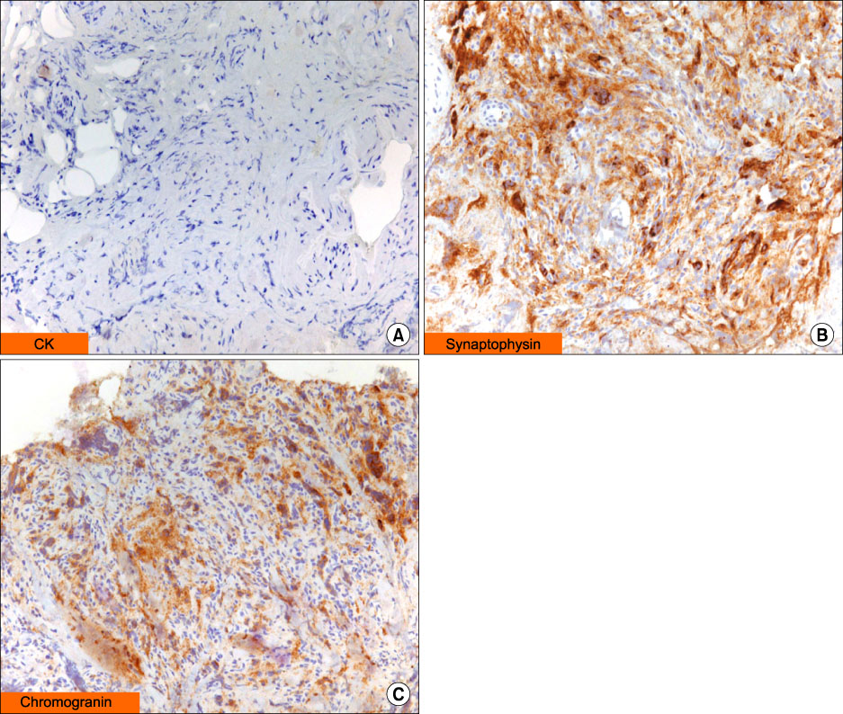
Figure 2 (A) The result of IHC staining for cytokeratin is negative (×40). (B) The results of IHC staining for synaptophysin (positive) (×200). (C) The results of IHC staining for chromogranin A (positive) (×100). All results are highly suggestive of paraganglioma.
Reference
-
1. Adler JT, Meyer-Rochow GY, Chen H, Benn DE, Robinson BG, Sippel RS, et al. Pheochromocytoma: current approaches and future directions. Oncologist. 2008. 13:779–793.2. Joynt KE, Moslehi JJ, Baughman KL. Paragangliomas: etiology, presentation, and management. Cardiol Rev. 2009. 17:159–164.3. Plouin PF, Gimenez-Roqueplo AP. Pheochromocytomas and secreting paragangliomas. Orphanet J Rare Dis. 2006. 1:49.4. Lee JA, Duh QY. Sporadic paraganglioma. World J Surg. 2008. 32:683–687.5. Young WF Jr. Paragangliomas: clinical overview. Ann NY Acad Sci. 2006. 1073:21–29.6. Scholz T, Schulz C, Klose S, Lehnert H. Diagnostic management of benign and malignant pheochromocytoma. Exp Clin Endocrinol Diabetes. 2007. 115:155–159.7. Petri BJ, van Eijck CH, de Herder WW, Wagner A, de Krijger RR. Phaeochromocytomas and sympathetic paragangliomas. Br J Surg. 2009. 96:1381–1392.8. Havekes B, Lai EW, Corssmit EP, Romijn JA, Timmers HJ, Pacak K. Detection and treatment of pheochromocytomas and paragangliomas: current standing of MIBG scintigraphy and future role of PET imaging. Q J Nucl Med Mol Imaging. 2008. 52:419–429.9. Moran CA, Suster S, Fishback N, Koss MN. Mediastinal paragangliomas. A clinicopathologic and immunohistochemical study of 16 cases. Cancer. 1993. 72:2358–2364.10. Gallivan MV, Chun B, Rowden G, Lack EE. Intrathoracic paravertebral malignant paraganglioma. Arch Pathol Lab Med. 1980. 104:46–51.11. Kim Y, Park SY, Ko YG, Ha JW, Chung N, Chang BC, et al. A case of intrapericardial pheochromocytoma. Korean J Med. 2006. 70:434–438.12. Chrisoulidou A, Kaltsas G, Ilias I, Grossman AB. The diagnosis and management of malignant phaeochromocytoma and paraganglioma. Endocr Relat Cancer. 2007. 14:569–585.13. Lloyd RV, Sisson JC, Shapiro B, Verhofstad AA. Immunohistochemical localization of epinephrine, norepinephrine, catecholamine-synthesizing enzymes, and chromogranin in neuroendocrine cells and tumors. Am J Pathol. 1986. 125:45–54.14. McNicol AM. Histopathology and immunohistochemistry of adrenal medullary tumors and paragangliomas. Endocr Pathol. 2006. 17:329–336.15. Tischler AS. Pheochromocytoma and extra-adrenal paraganglioma: updates. Arch Pathol Lab Med. 2008. 132:1272–1284.
- Full Text Links
-
- Actions
-
Cited
- CITED
-
- Close
- Share
-
- Similar articles
-
- A Case Report of Resection of a Mediastinal Paraganglioma: Why All the Fuss?
- Mediastinal Paraganglioma: Complete Resection Using Video-Assisted Thoracoscopic Surgery
- A case of Cushing's syndrome in ACTH-secreting mediastinal paraganglioma
- A Case of Recurred Paraganglioma of the Anterior Mediastinum A Case of Recurred Paraganglioma of the Anterior Mediastinum: A Case Report
- Anesthetic Experience for Resection of Posterior Mediastinal Paraganglioma: A case report
