Giant Cauda Equina Schwannoma with Dystrophic Calcifications : Case Report and Review of the Literature
- Affiliations
-
- 1Department of Neurosurgery, Spine Center, Seoul National University Bundang Hospital, Seoul National University College of Medicine, Seongnam, Korea.
- 2Department of Neurosurgery, Asan Medical Center, University of Ulsan College of Medicine, Seoul, Korea. scrhim@amc.seoul.kr
- KMID: 1441420
- DOI: http://doi.org/10.3340/jkns.2012.51.2.105
Abstract
- Giant spinal schwannoma of the cauda equine involving many nerve roots is rare, and ossification is usually not observed in the schwannoma. A 21-year-old man presented with a 12-month history of urinary dysfunction and numbness below the buttocks. Plain radiography showed scalloping of the posterior surface of the vertebral bodies from L4 to the sacrum, and magnetic resonance imaging and computed tomography revealed a giant cauda equina tumor with dystrophic calcification. The tumor was completely removed, with intraoperative neurophysiologic monitoring. Histopathologic examination showed that the tumor was a schwannoma. The patient's postoperative course was uneventful, with urinary function and numbness gradually improving. Although a giant schwannoma accompanied by dystrophic calcification is extremely rare, such a tumor can be removed safely and completely by meticulous dissection and careful neuromonitoring of the cauda equina spinal nerves involved in the tumor.
MeSH Terms
Figure
-
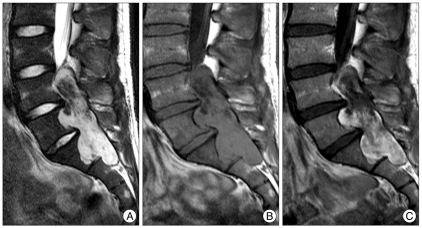
Fig. 1 Preoperative sagittal magnetic resonance images of our patient. A : T2-weighted image. B : T1-weighted image. C : Gadolinium-enhanced T1-weighted image. Note the giant cauda equina tumor growing into the vertebral bodies and neural foramina from L3 to S2.

Fig. 2 CT scans showing a large calcified mass in the enlarged spinal canal and neural foramen.

Fig. 3 Intraoperative photograph showing a large calcification (arrow) in the tumor.

Fig. 4 Radiographic images taken 3 years after surgery. A : T2-weighted magnetic resonance (MR) image. B : Gadolinium-enhanced T1-weighted MR image. C : Plain radiograph anteroposterior view. D : Plain radiograph lateral view. There is no evidence of any residual tumor, vertebral fracture, or spinal instability.
Reference
-
1. Bursztyn EM, Prada A. Intradural cauda equina schwannoma. Surg Neurol. 1986; 26:567–570. PMID: 3775634.
Article2. Cervoni L, Celli P, Scarpinati M, Cantore G. Neurinomas of the cauda equina clinical analysis of 40 surgical cases. Acta Neurochir (Wien). 1994; 127:199–202. PMID: 7942203.
Article3. Dickson JH, Waltz TA, Fechner RE. Intraosseous neurilemoma of the third lumbar vertebra. J Bone Joint Surg Am. 1971; 53:349–355. PMID: 5546707.
Article4. Hung CH, Tsai TH, Lieu AS, Lin CL, Lee KS, Hwang SL, et al. Giant invasive schwannoma of cauda equina with minimal neurologic deficit : a case report and literature review. Kaohsiung J Med Sci. 2008; 24:212–217. PMID: 18424359.
Article5. Jeon JH, Hwang HS, Jeong JH, Park SH, Moon JG, Kim CH. Spinal schwannoma; analysis of 40 cases. J Korean Neurosurg Soc. 2008; 43:135–138. PMID: 19096620.
Article6. Kagaya H, Abe E, Sato K, Shimada Y, Kimura A. Giant cauda equina schwannoma. A case report. Spine (Phila Pa 1976). 2000; 25:268–272. PMID: 10685494.7. Kim P, Ebersold MJ, Onofrio BM, Quast LM. Surgery of spinal nerve schwannoma. Risk of neurological deficit after resection of involved root. J Neurosurg. 1989; 71:810–814. PMID: 2585070.8. Kotoura Y, Shikata J, Yamamuro T, Kasahara K, Iwasaki R, Nakashima Y, et al. Radiation therapy for giant intrasacral schwannoma. Spine (Phila Pa 1976). 1991; 16:239–242. PMID: 2011787.
Article9. Lesoin F, Krivosic I, Cama A, Jomin M. A giant intrasacral schwannoma revealed by lumbosacral pain. Neurochirurgia (Stuttg). 1984; 27:23–24. PMID: 6700817.
Article10. Natarajan M, Rajagopal T, Srinivasan K. A giant schwannoma of cauda equina. Surg Neurol. 1975; 4:367–368. PMID: 1179257.11. Ortolan EG, Sola CA, Gruenberg MF, Carballo Vazquez F. Giant sacral schwannoma. A case report. Spine (Phila Pa 1976). 1996; 21:522–526. PMID: 8658260.12. Osborn RE, DeWitt JD. Giant cauda equina schwannoma : CT appearance. AJNR Am J Neuroradiol. 1985; 6:835–836. PMID: 3933309.13. Rengachary SS, O'Boynick P, Batnitzky S, Kepes JJ. Giant intrasacral Schwannoma : case report. Neurosurgery. 1981; 9:573–577. PMID: 7322323.14. Saito T, Shimode M, Azuma S, Seichi A. Giant schwannoma of the cauda equina with dural ectasia : a case report. J Orthop Sci. 2004; 9:635–637. PMID: 16228684.15. Salvant JB Jr, Young HF. Giant intrasacral schwannoma : an unusual cause of lumbrosacral radiculopathy. Surg Neurol. 1994; 41:411–413. PMID: 8009417.
Article16. Santi MD, Mitsunaga MM, Lockett JL. Total sacrectomy for a giant sacral schwannoma. A case report. Clin Orthop Relat Res. 1993; 285–289. PMID: 8358930.17. Turgut M, Erkuş M. Giant schwannoma of the cauda equina : case report and review of the literature. Zentralbl Neurochir. 2008; 69:99–101. PMID: 18444220.
Article
