Hepatocellular Carcinoma in the Right Thigh without Primary Hepatic Lesion: A Case Report
- Affiliations
-
- 1Department of Radiology, College of Medicine, Yeungnam University, Daegu, Korea. jcchang@med.yu.ac.kr
- 2Department of Radiology, College of Medicine, University of Ulsan, Asan Medical Center, Seoul, Korea.
- KMID: 1439542
- DOI: http://doi.org/10.3348/jksr.2012.67.4.285
Abstract
- Hepatocellular carcinoma (HCC) is the most common primary malignant tumor of the liver. There are some reports of primary extrahepatic HCC. But there is no report of HCC presenting as primary thigh mass. We report a case of HCC as a palpable mass in the right thigh, without hepatic lesion. On an MRI, the mass showed non-specific signal intensity and was progressively and centripetally enhanced.
MeSH Terms
Figure
-
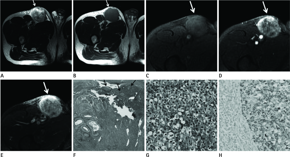
Fig. 1 A 56 years old man with palpable mass in the right thigh just below the inguinal area underwent pelvic MRI and the mass was surgically resected. A, B. The mass (arrows) is heterogeneous signal intensity with high signal intensity portion on T2 weighted image (A), and homogeneous intermediate to high signal intensity on T1 weighted image (B) when compared to those of thigh muscle. C-E. Dynamic contrast study shows progressively centripetal enhancement of the mass (arrows) on precontrast (C), arterial (D), and 5 minutes delayed (E) phases. F, G. Microscopically hepatoid tumor cells are seen and lymphoid follicles (arrows) are visible in the periphery of tumor cell clusters [hematoxylin-eosin stain, × 40 (F), × 400 (G)]. H. The tumor cells are positive for anti-human hepatocyte antibody (immunohistochemical stain, × 100).
Reference
-
1. Hong SS, Kim TK, Sung KB, Kim PN, Ha HK, Kim AY, et al. Extrahepatic spread of hepatocellular carcinoma: a pictorial review. Eur Radiol. 2003. 13:874–882.2. Longmaid HE 3rd, Seltzer SE, Costello P, Gordon P. Hepatocellular carcinoma presenting as primary extrahepatic mass on CT. AJR Am J Roentgenol. 1986. 146:1005–1009.3. Cho HG, Chung JP, Lee KS, Chon CY, Kang JK, Park IS, et al. Extrahepatic bile duct hepatocellular carcinoma without primary hepatic parenchymal lesions--a case report. Korean J Intern Med. 1996. 11:169–174.4. Uka K, Aikata H, Takaki S, Shirakawa H, Jeong SC, Yamashina K, et al. Clinical features and prognosis of patients with extrahepatic metastases from hepatocellular carcinoma. World J Gastroenterol. 2007. 13:414–420.5. Kummar S, Shafi NQ. Metastatic hepatocellular carcinoma. Clin Oncol (R Coll Radiol). 2003. 15:288–294.6. Katyal S, Oliver JH 3rd, Peterson MS, Ferris JV, Carr BS, Baron RL. Extrahepatic metastases of hepatocellular carcinoma. Radiology. 2000. 216:698–703.7. Shadbolt CL, Heinze SB, Dietrich RB. Imaging of groin masses: inguinal anatomy and pathologic conditions revisited. Radiographics. 2001. 21 Spec No:S261–S271.8. Wood AJ, Lappinga PJ, Ahmed I. Hepatocellular carcinoma metastatic to skin: diagnostic utility of antihuman hepatocyte antibody in combination with albumin in situ hybridization. J Cutan Pathol. 2009. 36:262–266.9. Watanabe J, Nakashima O, Kojiro M. Clinicopathologic study on lymph node metastasis of hepatocellular carcinoma: a retrospective study of 660 consecutive autopsy cases. Jpn J Clin Oncol. 1994. 24:37–41.
- Full Text Links
-
- Actions
-
Cited
- CITED
-
- Close
- Share
-
- Similar articles
-
- A case of spontaneous hepatic rupture in a patient with primary hepatocellular carcinoma during the puerperium
- Hepatocellular Carcinoma in Primary Biliary Cirrhosis: A Case Report
- Cystic Hepatocellular carcinoma: a case report
- Primary Hepatic Tuberculosis Mimicking Hepatocelluar Carcinoma in Patient with Chronic Viral Hepatitis B and C
- Metastatic hepatocellular carcinoma in the maxilla and temporal bone: a rare case report
