Gastritis Cystica Polyposa in the Unoperated Stomach: A Case Report
- Affiliations
-
- 1Department of Radiology, Research Institute of Radiological Science, Severance Hospital, Yonsei University College of Medicine, Seoul, Korea. bellenina@yuhs.ac
- 2Department of Pathology, Severance Hospital, Yonsei University College of Medicine, Seoul, Korea.
- KMID: 1439536
- DOI: http://doi.org/10.3348/jksr.2012.67.4.253
Abstract
- Gastritis cystica polyposa (GCP) is an uncommon lesion that usually develops at the gastroenterostomy site. A 57-year-old man visited a hospital with a complaint of melena. He did not have any surgical history or past medical history. Endoscopy was performed to evaluate the cause of melena, and a polypoid cystic mass in the stomach was found on an endoscopy and endoscopic ultrasonography. The polypoid cystic mass did not show any enhancing solid portion on a computed tomography. The gastric lesion was conclusively confirmed as GCP through endoscopic submucosal dissection. We report a rare case of GCP that occurred in an unoperated stomach.
Figure
-
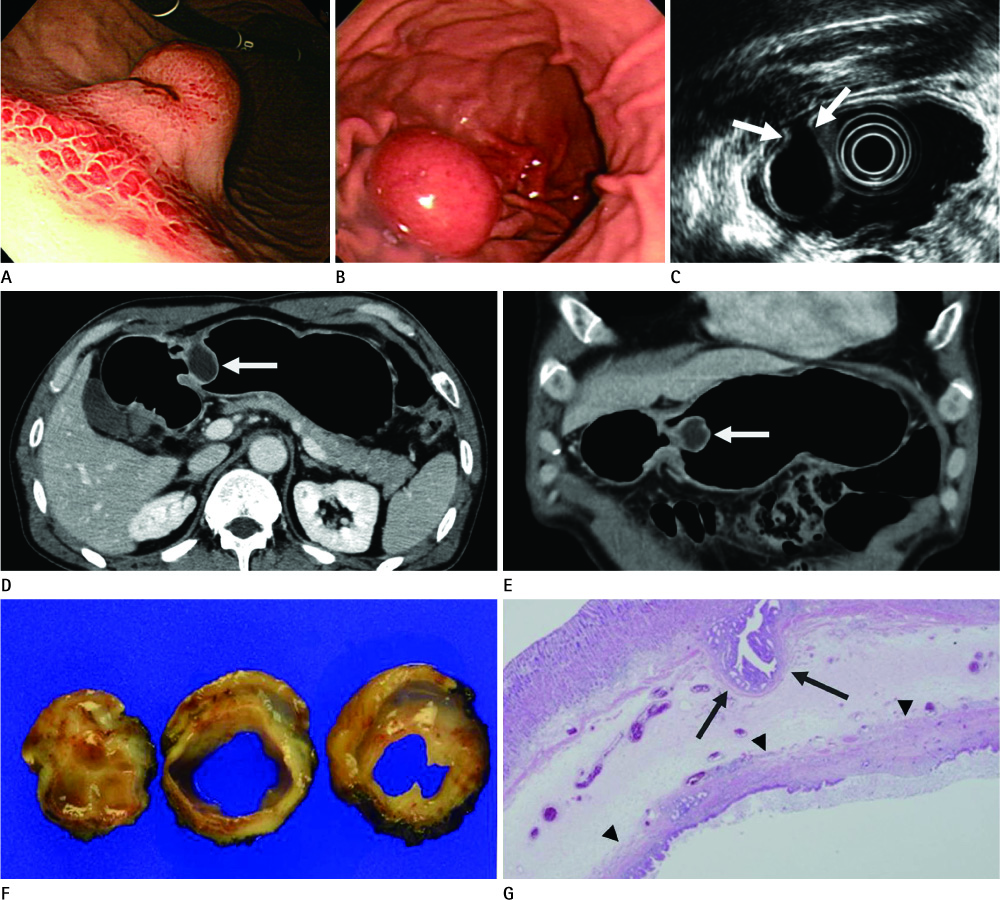
Fig. 1 A 57-year-old man with gastritis cystica polyposa in the unoperated stomach, which was confirmed by an endoscopic mucosal dissection. A. The initial endoscopic image shows a polypoid mass of 2.4 cm in size with mucosal ulceration and erythema on the mass. Submucosal hemorrhage is shown in the adjacent gastric mucosa. B. Erythema of gastric mucosa and ulceration on the surface of the polypoid mass are improved on the follow-up endoscopic image. C. EUS image shows a unilocular, cystic mass in the submucosal layer without any solid component. The mass has a narrow stalk (arrows). D, E. The axial (D) and coronal (E) images of contrast-enhanced CT show a polypoid, cystic mass (arrows) with a well-defined margin on the greater curvature of the stomach. Intact gastric mucosa is seen overlying the mass. F. The gross specimen shows a polypoid mass with a submucosal stroma measuring 2 × 1.8 × 2.5 cm. Cross-sectioning of the specimen reveals a unilocular cyst with a background of pale brown mucoid submucosal stroma. G. On microscopic examination, there is a large submucosal cyst (arrowheads) lined with gastric epithelium. Some elongated gastric glands (arrows) penetrate the muscularis mucosa (Hematoxylin-Eosin stain, × 12.5). Note.-EUS = endoscopic ultrasonography
Reference
-
1. Lee J, Park CM, Kim KA, Lee CH, Choi JW, Shin BK, et al. Cystic lesions of the gastrointestinal tract: multimodality imaging with pathologic correlations. Korean J Radiol. 2010. 11:457–468.2. Wu MT, Pan HB, Lai PH, Chang JM, Tsai SH, Wu CW. CT of gastritis cystica polyposa. Abdom Imaging. 1994. 19:8–10.3. Franzin G, Novelli P. Gastritis cystica profunda. Histopathology. 1981. 5:535–547.4. Béchade D, Desramé J, Algayres JP. Gastritis cystica profunda in a patient with no history of gastric surgery. Endoscopy. 2007. 39:Suppl 1. E80–E81.5. Tuncer K, Alkanat M, Musoğlu A, Aydin A. Gastritis cystica polyposa found in an unoperated stomach: an unusual case treated by endoscopic polypectomy. Endoscopy. 2003. 35:882.6. Littler ER, Gleibermann E. Gastritis cystica polyposa. (Gastric mucosal prolapse at gastroenterostomy site, with cystic and infiltrative epithelial hyperplasia). Cancer. 1972. 29:205–209.7. Park JS, Myung SJ, Jung HY, Yang SK, Hong WS, Kim JH, et al. Endoscopic treatment of gastritis cystica polyposa found in an unoperated stomach. Gastrointest Endosc. 2001. 54:101–103.8. Park CH, Park JM, Jung CK, Kim DB, Kang SH, Lee SW, et al. Early gastric cancer associated with gastritis cystica polyposa in the unoperated stomach treated by endoscopic submucosal dissection. Gastrointest Endosc. 2009. 69:e47–e50.9. Tomizuka T, Mazaki T, Mado K, Henmi A, Ishii Y, Masuda H, et al. A case of gastritis cystica profunda. Surgery. 2008. 143:449–450.
- Full Text Links
-
- Actions
-
Cited
- CITED
-
- Close
- Share
-
- Similar articles
-
- Gastritis Cystica Polyposa with Gastroduodenal Intussuception: A Case Report
- Gastritis Cystica Polyposa Arising from an Intact Stomach and Endoscopic Resection
- Endoscopically Treated Gastritis Cystica Polyposa Found in an Unoperated Stomach
- A Case of Gastritis Cystica Polyposa Presenting as Multiple Polypoid Lesions
- A Case of Gastritis Cystica Polyposa Presenting as a Hyperplastic Polyp
