Direct Communication with Fistula between the Left Main Pulmonary Artery and the Left Atrium: A Case Report
- Affiliations
-
- 1Department of Radiology, Chungnam National University Hospital, Chungnam National University School of Medicine, Daejeon, Korea. haneul88@hanmail.net
- 2Department of Cardiology, Chungnam National University Hospital, Chungnam National University School of Medicine, Daejeon, Korea.
- KMID: 1439419
- DOI: http://doi.org/10.3348/jksr.2012.66.3.235
Abstract
- We report a rare case of a direct communication-forming fistula between the left main pulmonary artery and left atrial appendage detected on computed tomography and color Doppler echocardiography.
Figure
-
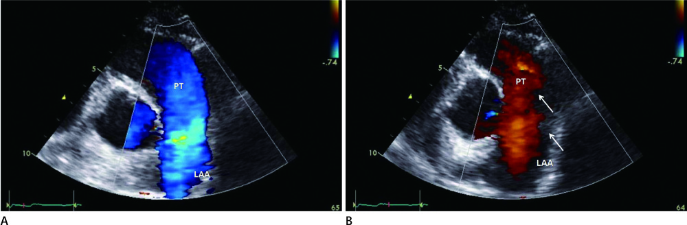
Fig. 1 A 23-year-old woman with a continuous cardiac murmur. A transthoracic color Doppler echocardiography was performed on the pulmonary trunk in systole (A) and diastole (B). Note the dilatation and regurgitant flow (red color flow, arrow) in the pulmonary trunk in diastole on the parasternal view Note.-PT = pulmonary trunk, LAA = left atrial appendage area
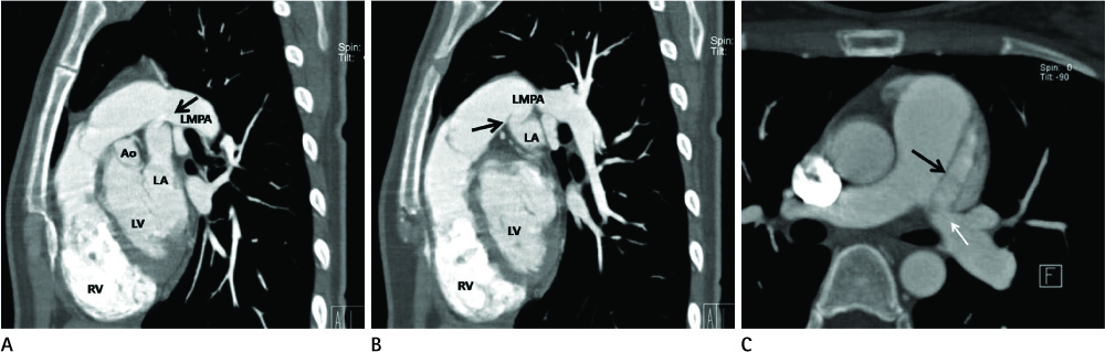
Fig. 2 A 23-year-old woman with a continuous cardiac murmur. Oblique maximum intensity projection (MIP) and axial images of computed tomography show an abnormal direct communication (arrow) from the inferior aspect of the LMPA, close to the bifurcation site of the pulmonary trunk, to the left atrial appendage (A-C). The maximum diameter of the communicating fistula vessel was 6 mm in size. Note.-Ao = ascending aorta, LMPA = left main pulmonary artery, LA = left atrium, LV = left ventricle, RV = right ventricle
Reference
-
1. Friedlich A, Bing RJ, Blount SG Jr. Physiological studies in congenital heart disease; circulatory dynamics in the anomalies of venous return to the heart including pulmonary arteriovenous fistula. Bull Johns Hopkins Hosp. 1950; 86:20–57.2. Mohanty SR, Yadav R, Kothari SS, Airan B. Right pulmonary artery left atrium communication. Ann Thorac Surg. 2000; 69:269–271.3. Lucas RV Jr, Lund GW, Edwards JE. Direct communication of a pulmonary artery with the left atrium. An unusual variant of pulmonary arteriovenous fistula. Circulation. 1961; 24:1409–1414.4. Orlick AE, Hultgren HN, Stoner JD, Barry WH, Wexler L, Dong EV Jr. Traumatic pulmonary artery--left atrial fistula: an unusual case of cyanosis in an adult. Am Heart J. 1979; 98:366–370.5. Karnik AM, Nilsson U, Vijayaraghavan G, Hashmi J, Shuhaiber H. Direct communication between the left pulmonary artery and the left atrium. Chest. 1989; 96:937–939.6. Faizal A, Madhu Sankar N, Murthy KS, Cherian KM. Right pulmonary artery-to-left atrial fistula. Asian Cardiovasc Thorac Ann. 2002; 10:80–82.7. Liu L, Wei X, Pan T. Congenital right pulmonary artery-to-left atrial fistula. Asian Cardiovasc Thorac Ann. 2010; 18:373–375.8. Ohara H, Ito K, Kohguchi N, Ohkawa Y, Akasaka T, Takarada M, et al. Direct communication between the right pulmonary artery and the left atrium. A case report and review of the literature. J Thorac Cardiovasc Surg. 1979; 77:742–747.9. Francis E, Sivakumar K, Kumar RK. Transcatheter closure of fistula between the right pulmonary artery and left atrium using the Amplatzer duct occluder. Catheter Cardiovasc Interv. 2004; 63:83–86.10. Batinica S, Gagro A, Bradić I, Marinović B. Congenital pulmonary arteriovenous fistula: a rare cause of cyanosis in childhood. Thorac Cardiovasc Surg. 1991; 39:105–106.
- Full Text Links
-
- Actions
-
Cited
- CITED
-
- Close
- Share
-
- Similar articles
-
- Surgical Treatment of an Aneurysmal Coronary Artery Fistula between the Left Coronary Artery and Right Atrium: A Case Report
- Successful Dual-Patch Closure of a Fistula between the Right Pulmonary Artery and the Left Atrium
- Giant Aneurysm of a Congenital Coronary Arteriovenous Fistula Arising from the Left Coronary Artery
- Congenital coronary artery fistula
- A Case of Coronary Arteriovenous Fistula Associated with Pulmonary Artery Aneurysm Confirmed by Multi Detector-Row Helical CT
