J Korean Soc Radiol.
2012 Mar;66(3):229-233. 10.3348/jksr.2012.66.3.229.
A New Formula to Estimate the Length of Right Upper Extremity Vein from Elbow Crease to Carina Calculated by Peripherally Inserted Central Catheter Insertion through Right Basilic Vein Puncture
- Affiliations
-
- 1Department of Radiology, Hallym University College of Medicine, Hallym University Sacred Heart Hospital, Anyang, Korea. jeyrad@hallym.or.kr
- 2Department of Radiology, Kangdong Sacred Heart Hospital, Hallym University College of Medicine, Seoul, Korea.
- 3Department of Occupational Medicine, Hallym University College of Medicine, Hallym University Sacred Heart Hospital, Anyang, Korea.
- KMID: 1439418
- DOI: http://doi.org/10.3348/jksr.2012.66.3.229
Abstract
- PURPOSE
To measure the length of the upper extremity vein between the elbow crease and the carina (elbow crease to carina length, ECL), to facilitate the appropriate positioning of the tip of the peripherally inserted central catheter (PICC).
MATERIALS AND METHODS
A total of 124 patients (64 men and 60 women; mean age 65.2 +/- 15.4 years; range, 21-90 years) inserted with PICC through the right basilic vein under fluoroscopy were included in this retrospective study. The ECL was determined as follows: ECL = (distance from elbow crease to puncture site) + (the catheter length of PICC) - (distance from carina to catheter tip on post-procedural chest radiograph). We analyzed the relationship between ECL and patient height.
RESULTS
The mean ECL through right basilic vein was 42.07 +/- 4.03 cm (27.5 to 52.2 cm). ECL was found to be significantly correlated with patient height: ECL (cm) = 0.24 x patient height (cm) + 3.75.
CONCLUSION
The formula developed in our study would be helpful for predicting the optimal catheter length during a blind bedside procedure of PICC via the right basilic vein.
MeSH Terms
Figure
-
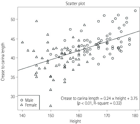
Fig. 1 Scatter plot of the length of the elbow crease to carina through the right basilic vein (cm) vs. patient height. The elbow crease to carina length (cm) = 0.24 × patient height (cm) + 3.75.
Cited by 1 articles
-
Outcomes of bedside peripherally inserted central catheter placement: a retrospective study at a single institution
Sukyung Kwon, Soo mi Son, Seul Hee Lee, Joung Hee Kim, Hyangkyoung Kim, Jang Yong Kim, Ji Il Kim, In Sung Moon
Acute Crit Care. 2020;35(1):31-37. doi: 10.4266/acc.2019.00731.
Reference
-
1. Ryder MA. Peripherally inserted central venous catheters. Nurs Clin North Am. 1993; 28:937–971.2. Bottino J, McCredie KB, Groschel DH, Lawson M. Long-term intravenous therapy with peripherally inserted silicone elastomer central venous catheters in patients with malignant diseases. Cancer. 1979; 43:1937–1943.3. Ng PK, Ault MJ, Ellrodt AG, Maldonado L. Peripherally inserted central catheters in general medicine. Mayo Clin Proc. 1997; 72:225–233.4. Kearns PJ, Coleman S, Wehner JH. Complications of long arm-catheters: a randomized trial of central vs peripheral tip location. JPEN J Parenter Enteral Nutr. 1996; 20:20–24.5. Tip of peripherally inserted central catheters. A position statement of the National Association of Vascular Access Networks. J Vasc Access Devices. 1998; 3:8–10.6. Lawson T. Infusion of IV medication and fluids via PICC and midline catheters: influences of tip position on the success of infusion. J Vasc Access Devices. 1998; 3:11–17.7. Fletcher SJ, Bodenham AR. Safe placement of central venous catheters: where should the tip of the catheter lie? Br J Anaesth. 2000; 85:188–191.8. Vesely TM. Central venous catheter tip position: a continuing controversy. J Vasc Interv Radiol. 2003; 14:527–534.9. Albrecht K, Nave H, Breitmeier D, Panning B, Tröger HD. Applied anatomy of the superior vena cava-the carina as a landmark to guide central venous catheter placement. Br J Anaesth. 2004; 92:75–77.10. Schuster M, Nave H, Piepenbrock S, Pabst R, Panning B. The carina as a landmark in central venous catheter placement. Br J Anaesth. 2000; 85:192–194.11. Yoon SZ, Shin JH, Hahn S, Oh AY, Kim HS, Kim SD, et al. Usefulness of the carina as a radiographic landmark for central venous catheter placement in paediatric patients. Br J Anaesth. 2005; 95:514–517.12. Stonelake PA, Bodenham AR. The carina as a radiological landmark for central venous catheter tip position. Br J Anaesth. 2006; 96:335–340.13. Neuman ML, Murphy BD, Rosen MP. Bedside placement of peripherally inserted central catheters: a cost-effectiveness analysis. Radiology. 1998; 206:423–428.14. James L, Bledsoe L, Hadaway LC. A retrospective look at tip location and complications of peripherally inserted central catheter lines. J Intraven Nurs. 1993; 16:104–109.15. Venkatesan T, Sen N, Korula PJ, Surendrababu NR, Raj JP, John P, et al. Blind placements of peripherally inserted antecubital central catheters: initial catheter tip position in relation to carina. Br J Anaesth. 2007; 98:83–88.16. Ragasa J, Shah N, Watson RC. Where antecubital catheters go: a study under fluoroscopic control. Anesthesiology. 1989; 71:378–380.17. Marino Paul L. Vascular access, peripherally inserted central catheters. The ICU book. 2007. 3rd ed. Lippincott Williams & Wilkins;p. 116.18. Lum P. A new formula-based measurement guide for optimal positioning of central venous catheters. JAVA. 2004; 9:80–85.19. Czepizak CA, O'Callaghan JM, Venus B. Evaluation of formulas for optimal positioning of central venous catheters. Chest. 1995; 107:1662–1664.
- Full Text Links
-
- Actions
-
Cited
- CITED
-
- Close
- Share
-
- Similar articles
-
- Simplified equation for determining proper depth of peripherally inserted central catheter in relation to anatomical landmarks
- Safety and Feasibility of Ultrasound-guided Peripherally Inserted Central Catheterization for Chemo-Delivery
- Anatomical Structures to Be Concerned With During Peripherally Inserted Central Catheter Procedures
- Placement of Peripherally Inserted Central Catheters (PICC): The Upper Arm Approach
- Proper Depth of Percutaneous Central Venous Catheter via the Great Saphenous Vein for Very Low Birth Weight Infants: A Single-Center, Prospective Cohort Study
