Validity of palatal superimposition of 3-dimensional digital models in cases treated with rapid maxillary expansion and maxillary protraction headgear
- Affiliations
-
- 1Department of Orthodontics, College of Dentistry, Gangneung-Wonju National University, Gangneung, Korea. korth@gwnu.ac.kr
- 2Department of Orthodontics, Dentofacial Orthopedics and Pedodontics, Center for Dental and Craniofacial Sciences, Charite-Universitatsmedizin, Berlin, Germany.
- KMID: 1435398
- DOI: http://doi.org/10.4041/kjod.2012.42.5.235
Abstract
OBJECTIVE
The purpose of this study was to evaluate the validity of the 3-dimensional (3D) superimposition method of digital models in patients who received treatment with rapid maxillary expansion (RME) and maxillary protraction headgear.
METHODS
The material consisted of pre- and post-treatment maxillary dental casts and lateral cephalograms of 30 patients, who underwent RME and maxillary protraction headgear treatment. Digital models were superimposed using the palate as a reference area. The movement of the maxillary central incisor and the first molar was measured on superimposed cephalograms and 3D digital models. To determine whether any difference existed between the 2 measuring techniques, intra-class correlation (ICC) and Bland-Altman plots were analyzed.
RESULTS
The measurements on the 3D digital models and cephalograms showed a very high correlation in the antero-posterior direction (ICC, 0.956 for central incisor and 0.941 for first molar) and a moderate correlation in the vertical direction (ICC, 0.748 for central incisor and 0.717 for first molar).
CONCLUSIONS
The 3D model superimposition method using the palate as a reference area is as clinically reliable for assessing antero-posterior tooth movement as cephalometric superimposition, even in cases treated with orthopedic appliances, such as RME and maxillary protraction headgear.
Keyword
Figure
-

Figure 1 The coordinate system and cephalometric superimposition of the maxilla along the palatal plane registered at anterior nasal spine (ANS). The X-axis was defined as the line through the maxillary central incisor tip and the mesio-buccal cusp tip of the first molar on the initial cephalogram, and the Y-axis as a line perpendicular to X-axis through the Sella. PNS, posterior nasal spine.
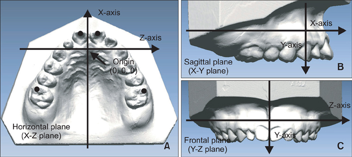
Figure 2 The coordinate system. A, The X-Z horizontal plane includes the origin, which is the junction of the incisive papilla and palatine raphe and is parallel to the occlusal plane constructed by bilateral mesio-buccal cusp tips of the first molars and the midpoint of the central incisors. B, The X-Y sagittal plane, which is perpendicular to the horizontal plane, is made up of the origin point and one arbitrary point on the mid-palatal suture. C, The Y-Z frontal plane is the section inclusive of the origin and perpendicular to both the sagittal and the horizontal planes. The measuring points were the midpoint on the edge of the upper central incisors and the mesio-buccal cusp tips of the upper first molars.

Figure 3 A, Superimposed models with differences displayed in colors on a millimeter scale. B, Superimposition of pre-treatment model (red) and post-treatment model (blue). Coronal view sectioned at C, the second premolars and D, the first molars.

Figure 4 Bland-Altman plots portraying the agreement between cephalometric and 3-dimensional (3D) measurements for A, antero-posterior tooth movement of central incisors; B, vertical tooth movement of central incisors; C, anteroposterior tooth movement of first molars; and D, vertical tooth movement of first molars. Each circle represents the difference between the measurements determined by the 2 methods (Y-axis) relative to the average of tooth movement measured by the 2 methods (X-axis). The thick lines indicate the mean, and the thin lines show the 95% limits of agreement.
Cited by 2 articles
-
A reliable method for evaluating upper molar distalization: Superimposition of three-dimensional digital models
Ruhi Nalcaci, Ayse Burcu Kocoglu-Altan, Ali Altug Bicakci, Firat Ozturk, Hasan Babacan
Korean J Orthod. 2015;45(2):82-88. doi: 10.4041/kjod.2015.45.2.82.Evaluation of adjacent tooth displacement in the posterior implant restoration with proximal contact loss by superimposition of digital models
Deuk-Won Jo, Min-Jung Kwon, Jong-Hee Kim, Young-Kyun Kim, Yang-Jin Yi
J Adv Prosthodont. 2019;11(2):88-94. doi: 10.4047/jap.2019.11.2.88.
Reference
-
1. Ricketts RM. A four-step method to distinguish orthodontic changes from natural growth. J Clin Orthod. 1975. 9:208–215. 218–228.2. Pancherz H. A cephalometric analysis of skeletal and dental changes contributing to Class II correction in activator treatment. Am J Orthod. 1984. 85:125–134.
Article3. Ghafari J, Baumrind S, Efstratiadis SS. Misinterpreting growth and treatment outcome from serial cephalographs. Clin Orthod Res. 1998. 1:102–106.
Article4. Peavy DC Jr, Kendrick GS. The effects of tooth movement on the palatine rugae. J Prosthet Dent. 1967. 18:536–542.
Article5. Almeida MA, Phillips C, Kula K, Tulloch C. Stability of the palatal rugae as landmarks for analysis of dental casts. Angle Orthod. 1995. 65:43–48.6. Bailey LT, Esmailnejad A, Almeida MA. Stability of the palatal rugae as landmarks for analysis of dental casts in extraction and nonextraction cases. Angle Orthod. 1996. 66:73–78.7. Hoggan BR, Sadowsky C. The use of palatal rugae for the assessment of anteroposterior tooth movements. Am J Orthod Dentofacial Orthop. 2001. 119:482–488.
Article8. Motohashi N, Kuroda T. A 3D computer-aided design system applied to diagnosis and treatment planning in orthodontics and orthognathic surgery. Eur J Orthod. 1999. 21:263–274.
Article9. Cha BK, Choi JI, Jost-Brinkmann PG, Jeong YM. Applications of three-dimensionally scanned models in orthodontics. Int J Comput Dent. 2007. 10:41–52.10. Lim MY, Lim SH. Comparison of model analysis measurements among plaster model, laser scan digital model, and cone beam CT image. Korean J Orthod. 2009. 39:6–17.
Article11. Lee SK, Kwon OW, Sung JH. A study on the dental arch characteristics of bialveolar protrusion patients using a three-dimensional digital model. Korean J Orthod. 2006. 36:45–54.12. Cha BK, Lee JY, Jost-Brinkmann PG, Yoshida N. Analysis of tooth movement in extraction cases using three-dimensional reverse engineering technology. Eur J Orthod. 2007. 29:325–331.
Article13. Commer P, Bourauel C, Maier K, Jager A. Construction and testing of a computer-based intraoral laser scanner for determining tooth positions. Med Eng Phys. 2000. 22:625–635.
Article14. Hayashi K, Araki Y, Uechi J, Ohno H, Mizoguchi I. A novel method for the three-dimensional (3-D) analysis of orthodontic tooth movement-calculation of rotation about and translation along the finite helical axis. J Biomech. 2002. 35:45–51.
Article15. Jang I, Tanaka M, Koga Y, Iijima S, Yozgatian JH, Cha BK, et al. A novel method for the assessment of three-dimensional tooth movement during orthodontic treatment. Angle Orthod. 2009. 79:447–453.
Article16. Miller RJ, Kuo E, Choi W. Validation of Align Technology's Treat III digital model superimposition tool and its case application. Orthod Craniofac Res. 2003. 6:Suppl 1. 143–149.
Article17. Ashmore JL, Kurland BF, King GJ, Wheeler TT, Ghafari J, Ramsay DS. A 3-dimensional analysis of molar movement during headgear treatment. Am J Orthod Dentofacial Orthop. 2002. 121:18–29.
Article18. van der Linden FP. Changes in the position of posterior teeth in relation to ruga points. Am J Orthod. 1978. 74:142–161.
Article19. Cha BK, Lee JY, Bae SH, Park DI. Preliminary study of future orthodontic model analysis: the orthodontic application of 3-dimensional reverse engineering technologies. J Korean Dent Assoc. 2002. 40:107–117.20. Friel S. Migration of teeth. Dent Rec (London). 1949. 69:74–84.21. Simmons JD, Moore RN, Erickson LC. A longitudinal study of anteroposterior growth changes in the palatine rugae. J Dent Res. 1987. 66:1512–1515.
Article22. Geran RG, McNamara JA Jr, Baccetti T, Franchi L, Shapiro LM. A prospective long-term study on the effects of rapid maxillary expansion in the early mixed dentition. Am J Orthod Dentofacial Orthop. 2006. 129:631–640.
Article23. Phatouros A, Goonewardene MS. Morphologic changes of the palate after rapid maxillary expansion: a 3-dimensional computed tomography evaluation. Am J Orthod Dentofacial Orthop. 2008. 134:117–124.
Article24. Schwarze CW. Does removal of the tooth germs of the third molars have an influence on the late form of the dental arch? Fortschr Kieferorthop. 1973. 34:387–400.25. Spillane LM, McNamara JA Jr. Maxillary adaptation to expansion in the mixed dentition. Semin Orthod. 1995. 1:176–187.
Article26. Jafari A, Shetty KS, Kumar M. Study of stress distribution and displacement of various craniofacial structures following application of transverse orthopedic forces: a three-dimensional FEM study. Angle Orthod. 2003. 73:12–20.27. Matsumoto M, Yoshii O. A case report of the rapid expansion of the maxillary dental arch by opening the mid palatal suture. Nihon Kyosei Shika Gakkai Zasshi. 1968. 27:166–174.28. Timms DJ. Rapid maxillary expansion. 1981. Chicago: Quintessence.29. Wertz R, Dreskin M. Midpalatal suture opening: a normative study. Am J Orthod. 1977. 71:367–381.
Article30. Biederman W. Rapid correction of Class 3 malocclusion by midpalatal expansion. Am J Orthod. 1973. 63:47–55.
- Full Text Links
-
- Actions
-
Cited
- CITED
-
- Close
- Share
-
- Similar articles
-
- A posteroanterior cephalometric study on the change of maxilla by rapid palatal expansion
- Displacement and stress distribution of the maxillofacial complex during maxillary protraction using palatal plates: A three-dimensional finite element analysis
- A study on the effect of rapid maxillary expansion and its relapse
- Spatial changes of the maxillofacial complex following maxillary protraction of human dry skull
- Crowding with no posterior crossbite treatment byrapid palatal expansion
