Korean J Clin Microbiol.
2012 Dec;15(4):151-153. 10.5145/KJCM.2012.15.4.151.
An Unusual Feature of Malaria: Exflagellated Microgametes of Malarial Parasites in Human Peripheral Blood
- Affiliations
-
- 1Department of Clinical Pathology, Kyungpook National University School of Medicine, Daegu, Korea. leewk@knu.ac.kr
- KMID: 1431813
- DOI: http://doi.org/10.5145/KJCM.2012.15.4.151
Abstract
- Exflagellation of the malaria parasite microgametocyte usually occurs in the gut cavity of Anopheles mosquitoes following an infective blood meal. Exflagellation is a very rare event in human blood. Due to its rarity, the appearance of this structure in a peripheral blood smear will easily create a diagnostic dilemma. We report a case of malaria with exflagellated microgametes in human blood that was initially mistaken for a double infection of Plasmodium and another blood flagellate. The patient was a 29-year-old Parkistani man presenting with fluctuating fever accompanied by chills and fatigue for 4 days. Initial peripheral blood smear examination showed a number of Plasmodium ring forms, trophozoites, and gametocytes. Additionally, several filamentous structures resembling blood flagellates were seen. With these features, an initial diagnostic impression of combined infection of malaria and blood flagellate was made. Later, we determined that these structures resembling blood flagellates were exflagellated microgametes of malarial parasite. Therefore, the knowledge that exflagellation may appear in human blood with Plasmodium species infection and being more familiar with differentiation of the morphologic features of other species infection can prevent further possible misinterpretation.
Figure
-

Fig. 1 Peripheral blood smear examination revealed the presence of rings and gametocytes (Wright stain, ×1,000).
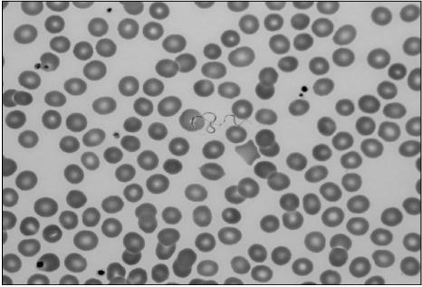
Fig. 2 Exflagellated microgametes in peripheral blood smear (Wright stain, ×1,000).
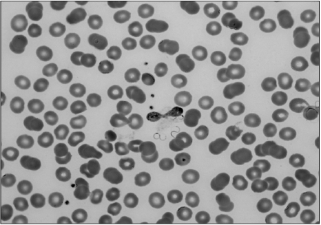
Fig. 3 Exflagellation of microgametes from microgametocytes in peripheral blood smear (Wright stain, ×1,000).
Reference
-
1. Gerber JE, Ukena TE, Cote L, Wyllie JM, Winn WC Jr. Exflagellation of malarial parasites in human peripheral blood. J Clin Microbiol. 1981. 13:236–237.2. Garnham PCC. Malaria Parasites and Other Hemosporidia. Oxford: Blackwell Scientific;1966–1114.3. Knell AJ. Knell AJ, editor. Malaria: The Problem. Malaria. 1991. Oxford: Oxford University Press;12–15.4. Carter R, Nijhout MM. Control of gamete formation (exflagellation) in malaria parasites. Science. 1977. 195:407–409.5. Prasad CS, Aparna N, Harendra Kumar ML. Exflagellated microgametes of Plasmodium vivax in human peripheral blood: an uncommon feature of malaria. Indian J Hematol Blood Transfus. 2011. 27:104–106.6. Enger A, Strand ØA, Ranheim T, Hellum KB. Exflagellation of microgametocytes in Plasmodium vivax malaria: a diagnostic conundrum. Med Princ Pract. 2004. 13:298–300.7. Tembhare P, Shirke S, Subramanian PG, Sehgal K, Gujral S. Exflagellated microgametes of Plasmodium vivax in human peripheral blood: a case report and review of the literature. Indian J Pathol Microbiol. 2009. 52:252–254.8. Ford JC, Wadsworth LD. Exflagellating Plasmodium vivax in peripheral blood. Arch Pathol Lab Med. 2003. 127:117–118.
- Full Text Links
-
- Actions
-
Cited
- CITED
-
- Close
- Share
-
- Similar articles
-
- A Case of Plasmodium vivax Infection Diagnosed after Treatment of Imported Falciparum Malaria
- Appearance Characteristics of the Atypical Lymphocytes in the Blood of the Plasmodium vivax Malarial Patients
- Two Cases of Malarial Infection in Pregnancy
- Case of Malarial Hepatitis by Plasmodium Vivax
- A relapsed case of imported tertian malaria after a standard course of hydroxychloroquine and primaquine therapy
