Intracranial Aneurysm Following Cranial Radiation Therapy
- Affiliations
-
- 1Department of Neurosurgery, Seoul National University Bundang Hospital, Department of Neurosurgery, Seoul National University College of Medicine, Seoul, Korea. nsbang@snubh.org
- KMID: 1431284
- DOI: http://doi.org/10.7461/jcen.2012.14.4.300
Abstract
- We report herein a case of a radiation-induced aneurysm. A 69-year-old woman presented with subarachnoid hemorrhage. Eight years previously, she had undergone cranial radiation therapy (total dose of 59.4 Gy) as adjuvant therapy after surgical resection for a chondrosarcoma that was destroying her sphenoid sinus. The patient underwent catheter angiography, which revealed an aneurysm of the anterior communicating artery and luminal narrowing and irregularity in the petrous and lacerum segments of the right internal carotid artery. We attempted surgical clipping of the aneurysm, but there was repeated bleeding. Finally the aneurysm was treated with endovascular trapping. Potentially fatal bleeding also occurred from her internal carotid artery, which had also been irradiated during the previous cranial radiation therapy. We stopped the bleeding with endovascular coil embolization. Because of diffuse vascular changes of the cerebral vessels within irradiated fields, special attention must be paid to their treatment.
MeSH Terms
Figure
-
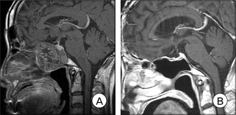
Fig. 1 A 69-year-old woman was found to have a large mass destroying her sphenoid sinus when she lost vision in her right eye eight years prior to current presentation (A). After treatment she was clinically monitored throughout the next eight years with serial Magnetic Resonance Imaging (MRI) and none of the MRI shows any sign of residual or recurrent tumor (B).
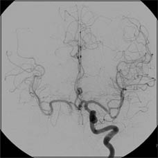
Fig. 2 Balloon occlusion test, which was performed before tumor removal, shows a good cross-filling via the anterior communicating and no vascular lesion.
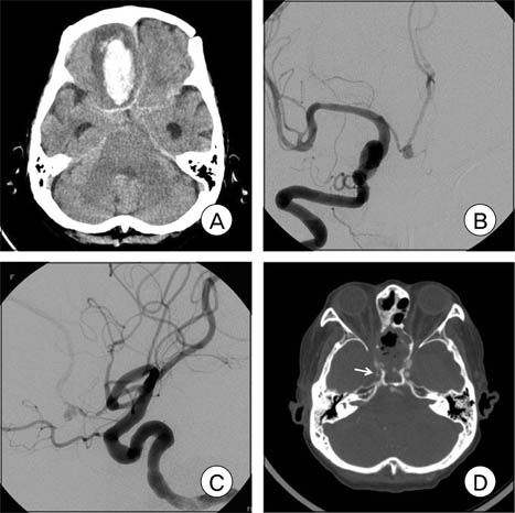
Fig. 3 Brain computed tomography (CT) shows intracerebral and subarachnoid hemorrhage (A). Angiography reveals an aneurysm of the anterior communicating artery (B and C). A source image of CT angiography shows luminal narrowing and irregularity in the petrous and lacerum segments of the right internal carotid artery (D).
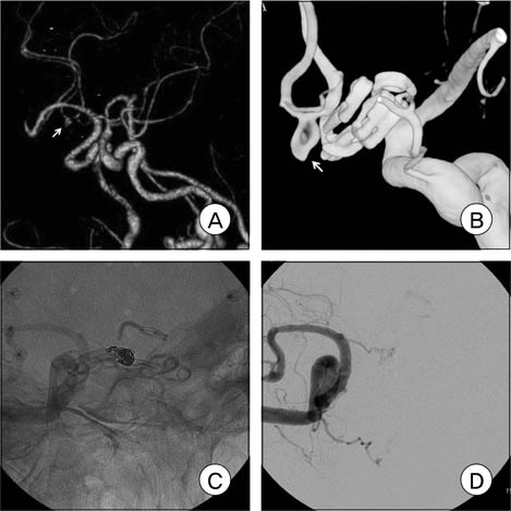
Fig. 4 CT angiography performed ten days after surgery reveals the small, residual neck of the aneurysm (A). Angiography which was performed ten days after clipping shows slippage of clips from the aneurysm (B). Postembolization angiograms (C and D) reveals successful obliteration of the aneurysm.

Fig. 5 Angiography shows contrast leakage from the lacerum segment of the right internal carotid artery into the nasal cavity, suggesting carotid blow out syndrome (A). After coil embolization in the ruptured point, no more leakage is noted on angiography (B).
Reference
-
1. Rowland JH, Hewitt M, Ganz PA. Cancer survivorship: a new challenge in delivering quality cancer care. J Clin Oncol. 2006. 11. 24(32):5101–5104.
Article2. Fajardo LF. The pathology of ionizing radiation as defined by morphologic patterns. Acta Oncol. 2005. 44(1):13–22.
Article3. Halle M, Hall P, Tornvall P. Cardiovascular disease associated with radiotherapy: activation of nuclear factor kappa-B. J Intern Med. 2011. 05. 269(5):469–477.
Article4. Bowers DC, Liu Y, Leisenring W, McNeil E, Stovall M, Gurney JG, et al. Late-occurring stroke among long-term survivors of childhood leukemia and brain tumors: a report from the Childhood Cancer Survivor Study. J Clin Oncol. 2006. 11. 24(33):5277–5282.
Article5. Haddy N, Mousannif A, Tukenova M, Guibout C, Grill J, Dhermain F, et al. Relationship between the brain radiation dose for the treatment of childhood cancer and the risk of long-term cerebrovascular mortality. Brain. 2011. 05. 134(Pt 5):1362–1372.
Article6. Sciubba DM, Gallia GL, Recinos P, Garonzik IM, Clatterbuck RE. Intracranial aneurysm following radiation therapy during childhood for a brain tumor. Case report and review of the literature. J Neurosurg. 2006. 08. 105(2):134–139.7. Morris B, Partap S, Yeom K, Gibbs IC, Fisher PG, King AA. Cerebrovascular disease in childhood cancer survivors: A Children's Oncology Group Report. Neurology. 2009. 12. 73(22):1906–1913.
Article8. Dunn IF, Woodworth GF, Siddiqui AH, Smith ER, Vates GE, Day AL, et al. Traumatic pericallosal artery aneurysm: a rare complication of transcallosal surgery. Case report. J Neurosurg. 2007. 106(2):153–157.9. Liu B, Zhang XM, Li QL. [Application of covered stent in the treatment of radiation-induced common carotid artery bleeding to a patient with nasoparyngeal carcinoma: a case report]. Beijing Da Xue Xue Bao. 2009. 12. 41(6):707–709. Chinese.10. Ellens D, Hurley MC, Surdel D, Shaibani A, Pelzer H, Bendok BR. Radiotherapy-induced common carotid pseudoaneurysm presenting with initially occult upper airway hemorrhage and successfully treated by endovascular stent graft. Am J Otolaryngol. 2010. May-Jun. 31(3):195–198.
Article11. Yucesoy K, Feiz-Erfan I, Spetzler RF, Han PP, Coons S. Anterior communicating artery aneurysm following radiation therapy for optic glioma: report of a case and review of the literature. Skull Base. 2004. 08. 14(3):169–173.
Article
- Full Text Links
-
- Actions
-
Cited
- CITED
-
- Close
- Share
-
- Similar articles
-
- Traumatic Aneurysm in the Posterior Cranial Fossa: Case Report
- Resolution of Third Cranial Nerve Palsy Following Endovascular Treatment of the Posterior Communicating Artery Aneurysm
- Intracranial "De Novo" Aneurysms: Case Report
- The Neuro-ophthalmic Presentation of Intracranial Aneurysms
- Aneurysm of the Posteior Inferior Cerebellar Artery Associated with Arteriovenous Malformation: Case Report
