J Korean Soc Magn Reson Med.
2013 Mar;17(1):55-58. 10.13104/jksmrm.2013.17.1.55.
Persistent Proatlantal Artery in Magnetic Resonance Angiography: A Case Report
- Affiliations
-
- 1Department of Radiology, Keimyung University Dongsan Hospital, College of Medicine, Korea. hyukwonchang@korea.com
- 2Department of General Surgery, Keimyung University Dongsan Hospital, College of Medicine, Korea.
- KMID: 1426753
- DOI: http://doi.org/10.13104/jksmrm.2013.17.1.55
Abstract
- Persistent proatlantal artery (PPA) is a rare embryologically remnant carotico-vertebrobasilar anastomoses. There are two types of PPA according to embryological considerations, origin and anatomic course. Type I PPA usually originate from internal carotid artery and not traversing transverse foramen. Type II PPA traverses from external carotid artery to C1 transverse foramen. The PPA is usually found incidentally without clinical symptoms, but can be related to several clinically significant vascular lesions, such as hypoplastic vertebral artery, intracranial arteriovenous malformation and in a case of carotid endarterectomy or external carotid artery embolization. So, thorough understanding of this anomaly is needed and we report a case of type II PPA diagnosed by MR angiography.
Keyword
MeSH Terms
Figure
-
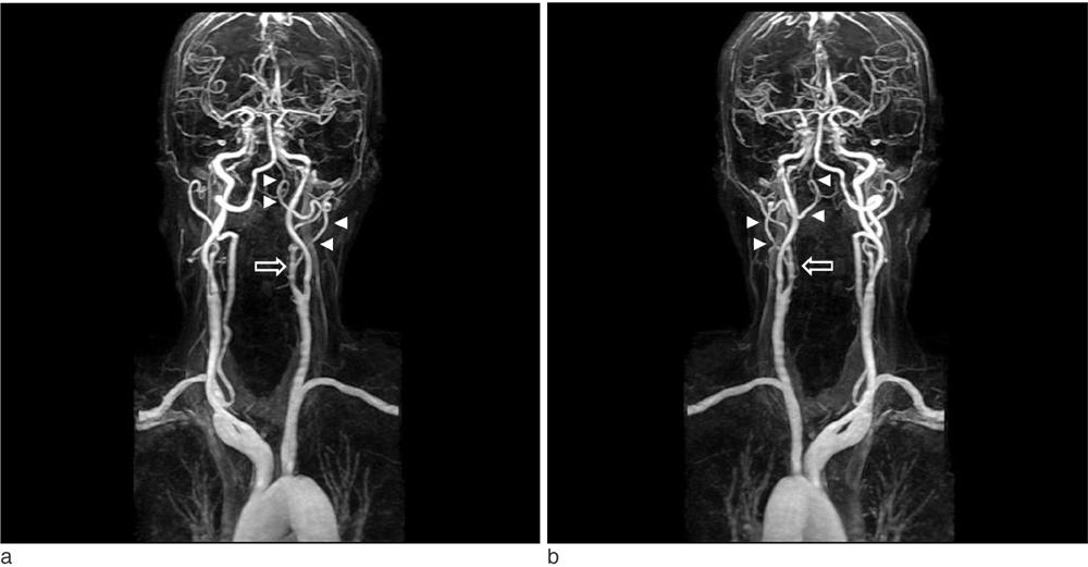
Fig. 1 Contrast-enhanced neck MR angiography. Left oblique view (a) and posterior-anterior view (b) shows Type II proatlantal artery which originate from left proximal ECA (arrow). This artery traverses from left proximal ECA to left vertebral artery ,V3 segment (arrow heads). There is aplasia of left vertebral artery involving V1 and V2 segments.
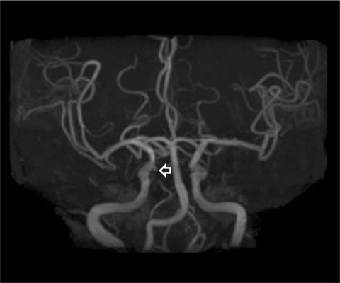
Fig. 2 Brain TOF (time-of flight) MR angiography shows about 2 mm sized small aneurysm of the right paraclinoid internal carotid artery (arrow).
Reference
-
1. Lasjaunias P, Theron J, Moret J. The occipital artery. Anatomy--normal arteriographic aspects--embryological significance. Neuroradiology. 1978; 15:31–33.2. Luh GY, Dean BL, Tomsick TA, Wallace RC. The persistent fetal carotid-vertebrobasilar anastomoses. AJR Am J Roentgenol. 1999; 172:1427–1432.3. Okahara M. Anatomic variations of the cerebral arteries and their embryology: a pictorial review. Eur Radiol. 2002; 12:2548–2561.4. Dimmick SJ, Faulder KC. Normal variants of the cerebral circulation at multidetector CT angiography. Radiographics. 2009; 29:1027–1043.5. Kwon JK, Kim MS, Lee CH. Persistent proatlantal artery Type I observed in a patient with subarachnoid hemorrhage: case report. Korean J Cerebrovasc Surg. 2008; 10:387–390.6. Yoo BG, Ji KT, Kim KS, Yoo KM, Kim SM, Joh YD. Proatlantal intersegmental artery Type II observed in a patient with lockedin syndrome. J Korean Neurol Assoc. 2002; 20:97–99.7. Gottschau M. Zwei seltene Varietäten der Stämme des aortenbogens. Arch Anat Entwicklgesch. 1885; 245–252.8. Padget DH. Designition of the embryonic intersegmental arteries in reference to the vertebral artery and subclavian stem. Anat Rec. 1954; 119:349–356.9. Arráez-Aybar LA, Navia-Alvarez P, Méndez-Cendón JC. A case of a type II proatlantal artery with arteriovenous malformation. Surg Radiol Anat. 2011; 33:85–89.10. Cohen JE, Grigoriadis S, Itshayek E. Type II proatlantal artery (occipital subtype) with bilateral absence of the vertebral arteries. Clin Anat. 2011; 24:950–952.
- Full Text Links
-
- Actions
-
Cited
- CITED
-
- Close
- Share
-
- Similar articles
-
- Persistent Proatlantal artery Type I Observed in a Patient with Subarachnoid Hemorrhage: Case Report
- A Type 1 Persistent Proatlantal Artery Originating from the External Carotid Artery Detected by Computed Tomographic Angiography
- Bilateral Agenesis of the Internal Carotid Artery: Case Report
- Persistent Trigeminal Artery Detected by Conventional Angiography and Magnetic Resonance Angiography
- Persistent Trigeminal Artery : An Unusual Cause of Cerebral Infarction
