A Novel Decorin Gene Mutation in Congenital Hereditary Stromal Dystrophy: A Korean Family
- Affiliations
-
- 1Department of Ophthalmology, Samsung Medical Center, Sungkyunkwan University School of Medicine, Seoul, Korea. tychung@skku.edu
- 2Department of Laboratory Medicine, Samsung Medical Center, Sungkyunkwan University School of Medicine, Seoul, Korea.
- KMID: 1387399
- DOI: http://doi.org/10.3341/kjo.2012.26.4.301
Abstract
- A 43-year-old man developed decreased vision in the right eye that had persisted for seven years. Under slit lamp examination, corneal clouding was noted with normal endothelium and ocular structure. From the clinical evidence, we suspected that the patient had congenital hereditary stromal dystrophy (CHSD). He and his family underwent a genetic analysis. Penetrating keratoplasty was conducted, and the corneal button was investigated for histopathologic confirmation via both light and electron microscopy. The histopathologic results revealed mildly loosened stromal structures, which exhibited an almost normal arrangement and differed slightly from the previous findings of CHSD cases. With regard to the genetic aspects, the patient and his mother harbored a novel point mutation of the decorin gene. This genetic mutation is also distinct from previously described deletion mutations of the decorin gene. This case involved delayed penetration of mild clinical symptoms with the histological feature of a loosened fiber arrangement in the corneal stroma. We concluded that this condition was a mild form of CHSD. However, from another perspective, this case could be considered as "decorin gene-associated corneal dystrophy," which is distinct from CHSD. Further evaluation will be required for appropriate clinical, histopathologic and genetic approaches for such cases.
MeSH Terms
Figure
-
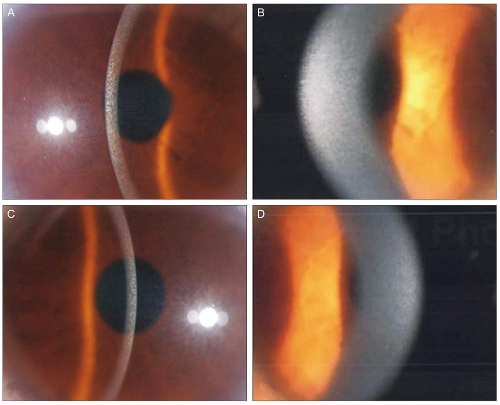
Fig. 1 Slit lamp photography of the patient. (A,B) Right eye. No gross abnormalities of the corneal endothelium, iris and lens were observed. Clouding of the cornea is noticeable under the arcuate slit beam. With magnification, ground-glass corneal opacities are more clearly seen in the anterior stroma, and identifiable small flakes and spots are present throughout the entire stoma. (C,D) Left eye. Density of corneal clouding is less than that of the right cornea.
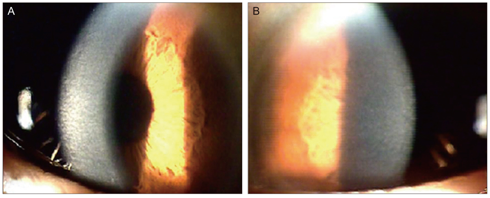
Fig. 2 Slit lamp photography of the patient's mother. (A) Right eye. Corneal stroma with arcuate slit beam shows diffuse clouding in the right eye. (B) Left eye. Ground-glass corneal opacities and small flakes are similar to that of the right eye.

Fig. 3 Pedigree of the family with stromal dystrophy. ▪ and • represent affected persons.
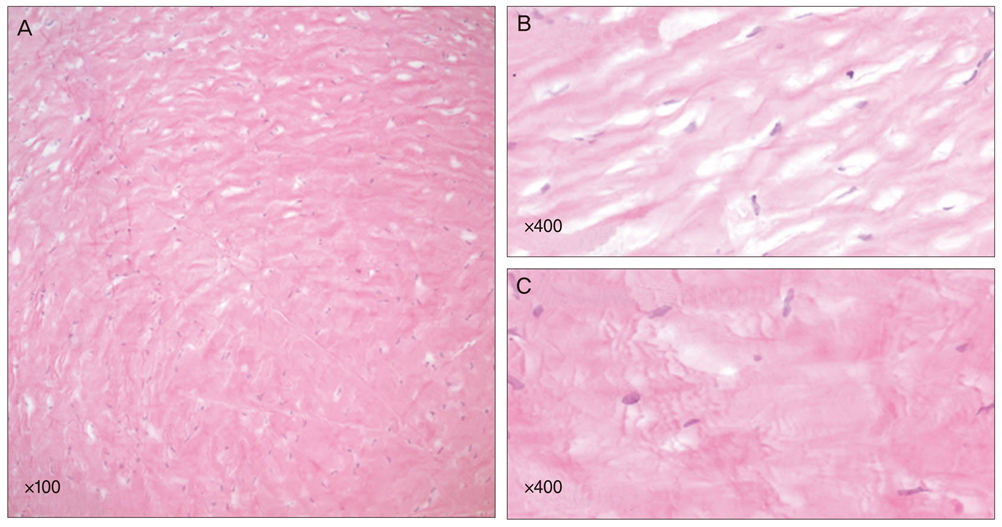
Fig. 4 Patient's light microscope findings. (A) Irregularity of corneal collagen fibril formation. Relatively loose arrangement of collagen fibers in the anterior stroma (B) and a relatively dense arrangement in the posterior stroma (C) are observed.
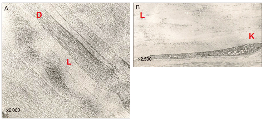
Fig. 5 Patient's transmission electron microscope findings. (A,B) A criss-crossing pattern of corneal fibers are shown with a relatively electron-dense (D) and lucent (L) structure. Irregularity in the collagen fibrils' shape and thickness is also seen. A keratocyte (K) is apparent in the electron lucent area (L).
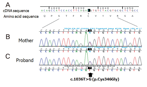
Fig. 6 Mutation analysis of the decorin gene. Partial sequence chromatograms displaying the wild-type DNA sequence of an unaffected person (A) and the DNA sequence of the mother (B) and patient (C), who were heterozygous for the decorin c.1036T point mutation.
Reference
-
1. Bredrup C, Knappskog PM, Majewski J, et al. Congenital stromal dystrophy of the cornea caused by a mutation in the decorin gene. Invest Ophthalmol Vis Sci. 2005. 46:420–426.2. Rodahl E, Van Ginderdeuren R, Knappskog PM, et al. A second decorin frame shift mutation in a family with congenital stromal corneal dystrophy. Am J Ophthalmol. 2006. 142:520–521.3. O'Connell JR, Weeks DE. PedCheck: a program for identification of genotype incompatibilities in linkage analysis. Am J Hum Genet. 1998. 63:259–266.4. Witschel H, Fine BS, Grutzner P, McTigue JW. Congenital hereditary stromal dystrophy of the cornea. Arch Ophthalmol. 1978. 96:1043–1051.5. Van Ginderdeuren R, De Vos R, Casteels I, Foets B. Report of a new family with dominant congenital heredity stromal dystrophy of the cornea. Cornea. 2002. 21:118–120.6. Bonfield JK, Rada C, Staden R. Automated detection of point mutations using fluorescent sequence trace subtraction. Nucleic Acids Res. 1998. 26:3404–3409.7. Michelacci YM. Collagens and proteoglycans of the corneal extracellular matrix. Braz J Med Biol Res. 2003. 36:1037–1046.8. Danielson KG, Baribault H, Holmes DF, et al. Targeted disruption of decorin leads to abnormal collagen fibril morphology and skin fragility. J Cell Biol. 1997. 136:729–743.
- Full Text Links
-
- Actions
-
Cited
- CITED
-
- Close
- Share
-
- Similar articles
-
- Congenital Herditary Stromal Dystrophy of the Cornea
- Two Cases of Congenital Hereditary Stromal Dystrophy of the Cornea
- N102S Mutation of UBIAD1 Gene in a Family with Schnyder Crystalline Corneal Dystrophy
- Infantile-Onset LMNA-Related Congenital Muscular Dystrophy Presenting as Torticollis: A Case Report
- A Case of Korean Patient with Macular Corneal Dystrophy Associated with Novel Mutation in the CHST6 Gene
