CT Angiographic Demonstration of a Mesenteric Vessel "Whirlpool" in Intestinal Malrotation and Midgut Volvulus: a Case Report
- Affiliations
-
- 1Gulhane Military Medical Academy, Department of Radiology, Ankara, Turkey. ubozlar@yahoo.com
- 2Gulhane Military Medical Academy, Turkish Armed Forces Rehabilitation Center, Division of Radiology, Ankara, Turkey.
- KMID: 1385407
- DOI: http://doi.org/10.3348/kjr.2008.9.5.466
Abstract
- Although the color Doppler ultrasonography diagnosis of intestinal malrotation with midgut volvulus, based on the typical "whirlpool" appearance of the mesenteric vascular structures is well-defined in the peer-reviewed literature, the combination of both the angiographic illustration of these findings and the contemporary state-of-the-art imaging techniques is lacking. We report the digital subtraction angiography and multidetector computed tomography angiography findings of a 37-year-old male with intestinal malrotation.
MeSH Terms
Figure
-
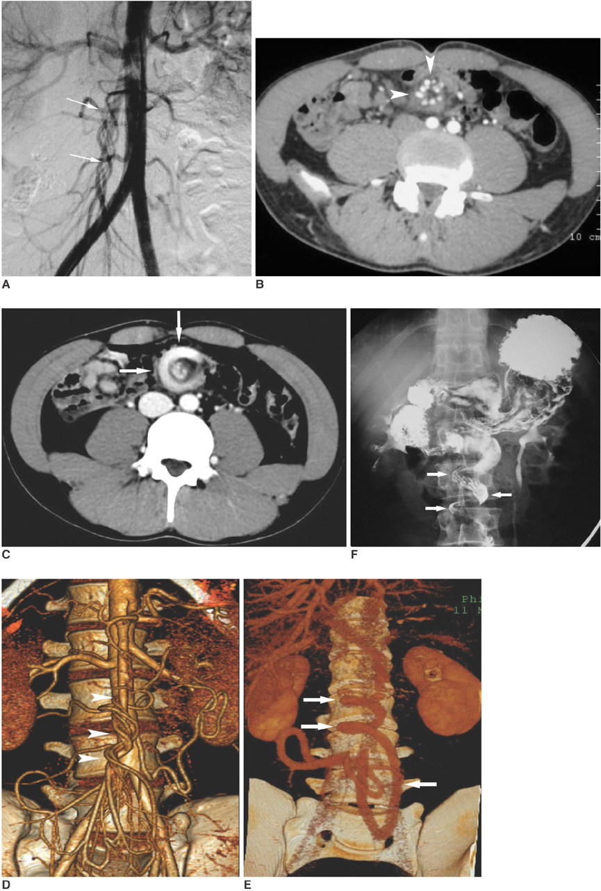
Fig. 1 Intestinal malrotation and midgut volvulus. A-F. Oblique digital subtraction angiogram (A) of patient depicting "barber's pole" sign (arrows) due to rotations of superior mesenteric artery and its branches. Axial CT (B, C) and 3D-MDCT angiography (D, E) images in arterial phase (B, D) and venous phase(C, E) depicting rotational abnormality of mesenteric arterial root and its branches (arrowheads, whirl sign) along with tortuous dilated superior mesenteric vein (arrows) which also contributes to "whirlpool" configuration. Note "clockwise" rotation (superior to inferior), as viewed from caudal aspect, of both arterial branches and mesenteric vein (at least partially on each other). Image from upper gastrointestinal series with barium (F), immediately after CT-angiography examination demonstrating "corkscrew" configuration (arrows) of proximal small bowel as it twists around superior mesenteric artery. Note that excreted renal pelvocalyceal contrast remaining from CT examination complicates this image.
Reference
-
1. Pracros JP, Sann L, Genin G, Tran-Minh VA, Morin de Finfe CH, Foray P, et al. Ultrasound diagnosis of midgut volvulus: the "whirlpool" sign. Pediatr Radiol. 1992. 22:18–20.2. Zerin JM, DiPietro MA. Superior mesenteric vascular anatomy at US in patients with surgically proved malrotation of the midgut. Radiology. 1992. 183:693–694.3. Taori K, Sanyal R, Attarde V, Bhagat M, Sheorain VS, Jawale R, et al. Unusual presentations of midgut volvulus with the whirlpool sign. J Ultrasound Med. 2006. 25:99–103.4. Buranasiri SI, Baum S, Nusbaum M, Tumen H. The angiographic diagnosis of midgut malrotation with volvulus in adults. Radiology. 1973. 109:555–556.5. Pickhardt PJ, Bhalla S. Intestinal malrotation in adolescents and adults: spectrum of clinical and imaging features. AJR Am J Roentgenol. 2002. 179:1429–1435.6. Zissin R, Rathaus V, Oscadchy A, Kots E, Gayer G, Shapiro-Feinberg M. Intestinal malrotation as an incidental finding on CT in adults. Abdom Imaging. 1999. 24:550–555.7. Berrocal T, Lamas M, Gutieerrez J, Torres I, Prieto C, del Hoyo ML. Congenital anomalies of the small intestine, colon, and rectum. Radiographics. 1999. 19:1219–1236.8. Long FR, Kramer SS, Markowitz RI, Taylor GE. Radiographic patterns of intestinal malrotation in children. Radiographics. 1996. 16:547–556.9. Clark P, Ruess L. Counterclockwise barber-pole sign on CT: SMA/SMV variance without midgut malrotation. Pediatr Radiol. 2005. 35:1125–1127.10. Fisher JK. Computed tomographic diagnosis of volvulus in intestinal malrotation. Radiology. 1981. 140:145–146.11. Bodard E, Monheim P, Machiels F, Mortelmans LL. CT of midgut malrotation presenting in an adult. J Comput Assist Tomogr. 1994. 18:501–502.12. Gollub MJ, Yoon S, Smith LM, Moskowitz CS. Does the CT whirl sign really predict small bowel volvulus?: Experience in an oncologic population? J Comput Assist Tomogr. 2006. 30:25–23.13. Weinberger E, Winters WD, Liddell RM, Rosenbaum DM, Krauter D. Sonographic diagnosis of intestinal malrotation in infants: importance of the relative positions of the superior mesenteric vein and artery. AJR Am J Roentgenol. 1992. 159:825–828.14. Orzech N, Navarro OM, Langer JC. Is ultrasonography a good screening test for intestinal malrotation? J Pediatr Surg. 2006. 41:1005–1009.15. Shimanuki Y, Aihara T, Takano H, Moritani T, Oguma E, Kuroki H, et al. Clockwise whirlpool sign at color Doppler US: an objective and definite sign of midgut volvulus. Radiology. 1996. 199:261–264.
- Full Text Links
-
- Actions
-
Cited
- CITED
-
- Close
- Share
-
- Similar articles
-
- Malrotation complicating Midgut Volvulus: Ultrasonographic Finding
- Intestinal Malrotation with Concurrent Portal Vein and Superior Mesenteric Vein Thromboses
- Midgut Volvulus in a 70-year-old Man Due to Intestinal Nonrotation
- A Case of Intestinal Malrotation Complicated by Midgut Volvulus: Diagnosis with Abdominal CT Scan
- Ileal Duplication in a Neonate With Jejuno-Ileal Atresia, Midgut Malrotation and Volvulus
