Soft Tissue Surgery for Equinus Deformity in Spastic Hemiplegic Cerebral Palsy: Effects on Kinematic and Kinetic Parameters
- Affiliations
-
- 1Department and Research Institute of Rehabilitation Medicine, Yonsei University College of Medicine, Seoul, Korea. medicus@yumc.yonsei.ac.kr
- 2Department of Orthopaedic Surgery, Yonsei University College of Medicine, Seoul, Korea.
- KMID: 1381246
- DOI: http://doi.org/10.3349/ymj.2006.47.5.657
Abstract
- The purpose of this study was to evaluate how soft tissue surgery for correcting equinus deformity affects the kinematic and kinetic parameters of the ankle and proximal joints. Sixteen children with spastic hemiplegic cerebral palsy and equinus deformities (age range 3-16 years) were included. Soft tissue surgeries were performed exclusively on the ankle joint area in all subjects. Using computerized gait analysis (Vicon 370 Motion Analysis System), the kinematic and kinetic parameters during barefoot ambulation were collected preoperatively and postoperatively. In all 16 children, the abnormally increased ankle plantar flexion and pelvis anterior tilting on the sagittal plane were significantly improved without a weakening of push-off (p < 0.05). In a group of 8 subjects with a recurvatum knee gait pattern before operation, the postoperative kinematic and kinetic parameters of the knee joint were significantly improved (p < 0.05). In a group of 8 subjects with ipsilateral pelvic external rotation before operation, the postoperative pelvic deviations on the transverse plane were significantly decreased (p < 0.05). These findings suggest that the soft tissue surgery for correcting equinus deformity improves not only the abnormal gait pattern of the ankle, but also that of the knee and pelvis.
Keyword
MeSH Terms
Figure
-

Fig. 1 Postoperative ankle kinematic and kinetic parameters on the sagittal plane in all 16 patients, as compared with preoperative and normal values. (A) ankle flexion (degrees), (B) ankle power generation (watts/kg). Dorsi, Dorsiflexion; Plantar, Plantarflexion; Gen, Generation; Abs, Absorption.

Fig. 2 Postoperative knee kinematic and kinetic parameters in 8 patients with recurvatum knee gait patterns, as compared with preoperative and normal values. (A) knee flexion (degrees), (B) knee moment (Nm/kg). Flex, Flexion; Ext, Extension.
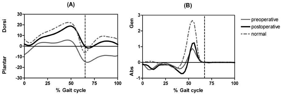
Fig. 3 Postoperative pelvic kinematic parameters on the transverse plane in 8 patients with the ipsilateral pelvic external rotation during gait cycle, as compared with preoperative and normal values. Int, Internal rotation; Ext, External rotation.
Cited by 1 articles
-
Botulinum Toxin Type A Injection for Spastic Equinovarus Foot in Children with Spastic Cerebral Palsy: Effects on Gait and Foot Pressure Distribution
Ja Young Choi, Soojin Jung, Dong-wook Rha, Eun Sook Park
Yonsei Med J. 2016;57(2):496-504. doi: 10.3349/ymj.2016.57.2.496.
Reference
-
1. Sutherland DH, Davids JR. Common gait abnormalities of the knee in cerebral palsy. Clin Orthop Relat Res. 1993. 288:139–147.2. Rodda JM, Graham HK, Carson L, Galea MP, Wolfe R. Sagittal gait patterns in spastic diplegia. J Bone Joint Surg Br. 2004. 86:251–258.3. Miller F, Dabney KW, Rang M. Epps CHJ, Bowen R, editors. Complications in cerebral palsy treatment. Complications in pediatric orthopaedic surgery. 1995. Philadelphia: JB Lippincott Company;477–544.4. Rang M, Silver R, De-La-Garza J. Lovell WW, Winter RB, editors. Cerebral palsy. Pediatric orthopaedics. 1986. Philadelphia: JB Lippincott Company;345–396.5. Aminian A, Vankoski SJ, Dias L, Novak RA. Spastic hemiplegic cerebral palsy and the femoral derotation osteotomy: effect at the pelvis and hip in the transverse plane during gait. J Pediatr Orthop. 2003. 23:314–320.6. Winters TF Jr, Gage JR, Hicks R. Gait patterns in spastic hemiplegia in children and young adults. J Bone Joint Surg Am. 1987. 69:437–441.7. Rose SA, DeLuca PA, Davis RB 3rd, Ounpuu S, Gage JR. Kinematic and kinetic evaluation of the ankle after lengthening of the gastrocnemius fascia in children with cerebral palsy. J Pediatr Orthop. 1993. 13:727–732.8. Steinwender G, Saraph V, Zwick EB, Uitz C, Linhart W. Fixed and dynamic equinus in cerebral palsy: evaluation of ankle function after multilevel surgery. J Pediatr Orthop. 2001. 21:102–107.9. Lyon R, Liu X, Schwab J, Harris G. Kinematic and kinetic evaluation of the ankle joint before and after tendo achilles lengthening in patients with spastic diplegia. J Pediatr Orthop. 2005. 25:479–483.10. Baddar A, Granata K, Damiano DL, Carmines DV, Blanco JS, Abel MF. Ankle and knee coupling in patients with spastic diplegia: Effects of gastrocnemiussoleus lengthening. J Bone Joint Surg Am. 2002. 84:736–744.11. Borton DC, Walker K, Pirpiris M, Nattrass GR, Graham HK. Isolated calf lengthening in cerebral palsy. Outcome analysis of risk factors. J Bone Joint Surg Br. 2001. 83:364–370.12. Chang CH, Albarracin JP, Lipton GE, Miller F. Long-term follow-up of surgery for equinovarus foot deformity in children with cerebral palsy. J Pediatr Orthop. 2002. 22:792–799.13. Gage JR, Ounpuu S. Pastla AE, editor. Surgical intervention in the correction of primary and secondary gait abnormalities. Adaptability of human gait. 1991. North Holland: Elsevier Science Publishers BV;359–385.14. Preiss RA, Condie DN, Rowley DI, Graham HK. The effects of botulinum toxin (BTX-A) on spasticity of the lower limb and on gait in cerebral palsy. J Bone Joint Surg Br. 2003. 85:943–948.15. Goodman MJ, Menown JL, West JM Jr, Barr KM, Vander Linden DW, McMulkin ML. Secondary gait compensations in individuals without neuromuscular involvement following a unilateral imposed equinus constraint. Gait Posture. 2004. 20:238–244.16. Perry J. Gait analysis; Normal and pathological function. 1992. 1st ed. Thorofare (NJ): SLACK Incorporated.17. Lin CJ, Guo LY, Su FC, Chou YL, Cherng RJ. Common abnormal kinetic patterns of the knee in gait in spastic diplegia of cerebral palsy. Gait Posture. 2000. 11:224–232.18. Perry J, Hoffer MM, Giovan P, Antonelli D, Greenberg R. Gait analysis of the triceps surae in cerebral palsy. A preoperative and postoperative clinical and electromyographic study. J Bone Joint Surg Am. 1974. 56:511–520.19. Gaines RW, Ford TB. A systematic approach to the amount of Achilles tendon lengthening in cerebral palsy. J Pediatr Orthop. 1984. 4:448–451.20. Grabe RP, Thompson P. Lengthening of the Achilles tendon in cerebral paresis: Basic principles and follow-up study. S Afr Med J. 1979. 56:993–996.21. Graham HK, Baker R, Dobson F, Morris ME. Multilevel orthopaedic surgery in group IV spastic hemiplegia. J Bone Joint Surg Br. 2005. 87:548–555.22. Saraph V, Zwick EB, Zwick G, Dreier M, Steinwender G, Linhart W. Effect of derotation osteotomy of the femur on hip and pelvis rotations in hemiplegic and diplegic children. J Pediatr Orthop B. 2002. 11:159–166.23. Noritake K, Stout JL, Gage JR. Pelvic rotation during walking in children with spastic hemiplegia cerebral palsy. Gait Posture. 1998. 7:164.
- Full Text Links
-
- Actions
-
Cited
- CITED
-
- Close
- Share
-
- Similar articles
-
- Changes in Gait Pattern After Surgeries for Equinus Gait in Cerebral Palsy Spastic Hemiplegia
- Kinematic and Kinetic Changes of the Ankle After the Correction of Spastic Equinus Deformity: Z-plastic Lengthening versus Strayer Method
- The Changes of Foot Pressure Distribution in Spastic Cerebral Palsy with Equinus Deformity following Corrective Surgery
- Management of recurred spastic equinus deformity by heel cord advancement in cerebral palsy
- Treatment of Calcaneovalgus Deformity Following Operative Treatment of Diplegic Equinovarus Deformity in Cerebral Palsy Patient: A Case Report
