Primary Uterine Lymphoma: A Case Report
- Affiliations
-
- 1Department of Diagnostic Radiology, Korea University Guro Hospital, Seoul, Korea.
- KMID: 1378950
- DOI: http://doi.org/10.3348/kjr.2000.1.4.223
Abstract
- Primary lymphoma of the uterus is a rare disease, the reported characteristic MR imaging findings being homogeneous intermediate signal intensity of the indistinct mass on T1- and T2-weighted images, and the preservation of endome-trial lining and uterine architecture. We report a case of primary uterine lymphoma which showed tumoral necrosis, endometrial disruption and diffuse anterior vagi-nal wall involvement.
MeSH Terms
Figure
-
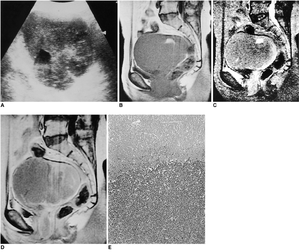
Fig. 1 Primary lymphoma in a 62-year-old woman. A. US reveals hypoechoic lobulated uterine enlargement, with an anechoic portion. An echogenic endometrial line is not present. B. Sagittal T1-weighted image shows homogeneous diffuse enlargement of the uterus, with an area of focal high signal intensity. The anterior vaginal wall is thickened. C. Sagittal T2-weighted image shows a low signal intensity uterus containing a high signal intensity area representing hemorrhagic necrosis. The zonal anatomy of the endometrium, myometrium and endocervix is not preserved, and the junctional zone is indistinct. D. T1-weighted image after gadolinium administration reveals inhomogeneous, moderate enhancement of the uterus and anterior vaginal wall. E. On photomicrograph the border between the myometrium and endometrium is blurred by the infiltration of lymphoid tumor cells, and the tumor shows extensive necrosis (H & E).
Reference
-
1. Komaki R, Cox J, Hansen R, Gunn W, Greeberg M. Malignant lymphoma of the uterus and cervix. Cancer. 1984. 54:1699–1974.2. Harri NL, Scully RE. Malignant lymphoma and granulocytic sarcoma of the uterus and vagina. Cancer. 1984. 3:2530–2545.3. Kawakami S, Togashi K, Kojima N, Morikawa K, Mori T, Konishi J. MR appearance of maliganant lymphoma of the uterus. J Comput Assist Tomogr. 1995. 19:238–242.4. Yamamda I. Primary uterine lymphoma: MR imaging. AJR. 1993. 160:662–663.5. Dang HT, Terk HR, Colleti PM, Schlaerth J, Cutin J. Primary lymphoma of the cervix: MRI findings with gadolinium. Magn Reson lmaging. 1991. 9:941–944.6. Kimura I, Togashi K, Tsutsui K, et al. MR imaging of gynecologic lymphoma. J Comput Assist Tomogr. 1991. 15:500–501.7. Kim YS, Joh BH, Cho OK, Rhim HC. MR imaging of primary uterine lymphoma. Abdom Imaging. 1997. 22:441–444.8. Stickler JG, Burgart LJ, Weiss LM. Weiss LM, editor. Classical Hodgkin's Disease. Pathology of lymph nodes: Contemporary issues in surgical patholgy. 1996. Churchill Livingstone;1–214.
